. 2000 May;74(10):4562-9.
doi: 10.1128/jvi.74.10.4562-4569.2000.

Multiple antiviral activities of cyanovirin-N: blocking of human immunodeficiency virus type 1 gp120 interaction with CD4 and coreceptor and inhibition of diverse enveloped viruses

Affiliations

Multiple antiviral activities of cyanovirin-N: blocking of human immunodeficiency virus type 1 gp120 interaction with CD4 and coreceptor and inhibition of diverse enveloped viruses

B Dey et al. J Virol. 2000 May.

Abstract

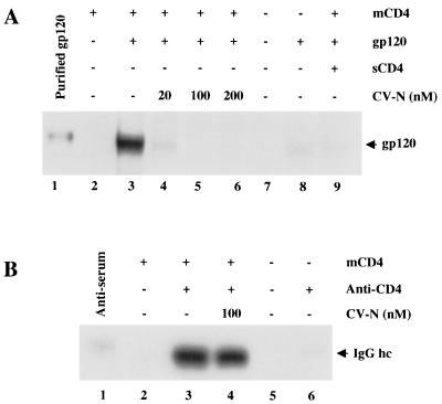
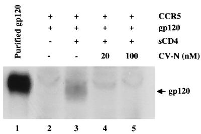
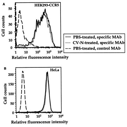
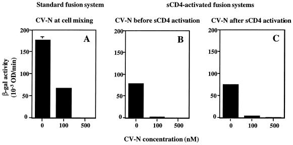
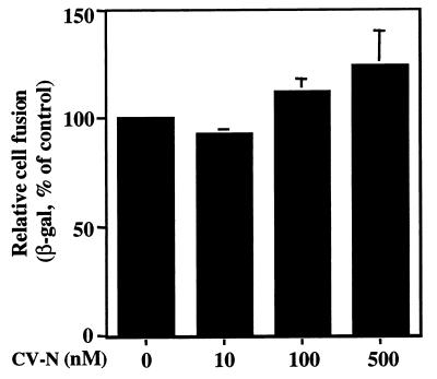
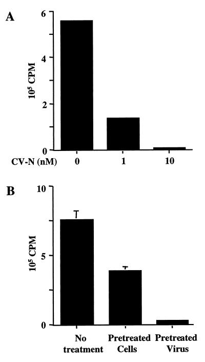
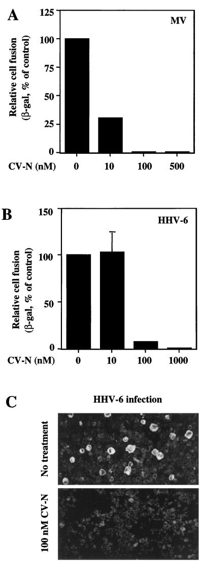
Cyanovirin-N (CV-N) is a cyanobacterial protein with potent neutralizing activity against human immunodeficiency virus (HIV). CV-N has been shown to bind HIV type 1 (HIV-1) gp120 with high affinity; moreover, it blocks the envelope glycoprotein-mediated membrane fusion reaction associated with HIV-1 entry. However, the inhibitory mechanism(s) remains unclear. In this study, we show that CV-N blocked binding of gp120 to cell-associated CD4. Consistent with this, pretreatment of gp120 with CV-N inhibited soluble CD4 (sCD4)-dependent binding of gp120 to cell-associated CCR5. To investigate possible effects of CV-N at post-CD4 binding steps, we used an assay that measures sCD4 activation of the HIV-1 envelope glycoprotein for fusion with CCR5-expressing cells. CV-N displayed equivalently potent inhibitory effects when added before or after sCD4 activation, suggesting that CV-N also has blocking action at the level of gp120 interaction with coreceptor. This effect was shown not to be due to CV-N-induced coreceptor down-modulation after the CD4 binding step. The multiple activities against the HIV-1 envelope glycoprotein prompted us to examine other enveloped viruses. CV-N potently blocked infection by feline immunodeficiency virus, which utilizes the chemokine receptor CXCR4 as an entry receptor but is CD4 independent. CV-N also inhibited fusion and/or infection by human herpesvirus 6 and measles virus but not by vaccinia virus. Thus, CV-N has broad-spectrum antiviral activity, both for multiple steps in the HIV entry mechanism and for diverse enveloped viruses. This broad specificity has implications for potential clinical utility of CV-N.


Figures

FIG. 1
FIG. 1
Effect of CV-N on binding of gp120 and anti-CD4 antibodies to cell-associated CD4. (A) Binding of mock-treated (lane 3) or CV-N-treated (lanes 4 to 6) JR-FL gp120 to CD4-expressing (vCB-3-infected) cells was determined by Western blotting of whole cell lysates using polyclonal anti-gp120 antibody for detection. For lane 9, gp120 was preincubated with excess of sCD4. As negative control, CD4-negative (WR-infected) cells were incubated without (lane 7) or with (lane 8) gp120. The indicated CV-N concentrations represent those in the final incubation mixtures. Purified JR-FL gp120 (lane 1) was run as positive standard for immunodetection. mCD4, membrane-associated CD4. (B) Effect of CV-N on binding of anti-CD4 polyclonal antiserum to CD4-expressing (lanes 2 to 4) or CD4-negative cells (lanes 5 and 6) was determined by Western blotting of whole cell lysates using horseradish peroxidase-conjugated goat anti-rabbit IgG. The indicated CV-N concentrations represent those in the final incubation mixture. In lane 1, diluted antiserum was run as a positive standard. Migration of IgG heavy chain (hc) is indicated.
FIG. 2
FIG. 2
Effect of CV-N on CD4-dependent gp120 binding to target cell coreceptor. Binding of mock-treated (lanes 2 and 3) or CV-N-treated (lanes 4 and 5) JR-FL gp120 to CCR5-expressing cells, upon activation by sCD4, was determined by Western blotting of whole cell lysates using polyclonal anti-gp120 antibody for detection. Lane 1 shows purified gp120 used as positive control for immunodetection. The indicated CV-N concentrations represent those in the final incubation mixtures. The faint band above gp120 in lanes 2 to 5 is a cellular protein that nonspecifically reacts with the antiserum.
FIG. 3
FIG. 3
Effect of CV-N on cell surface expression of HIV coreceptors. Cells were incubated with PBS alone or PBS plus 250 nM CV-N prior to staining with the indicated MAbs, followed by flow cytometric analysis. (A) For detection of CCR5, HEK293-CCR5 cells were stained with the specific MAb (2D7, PE conjugated); as a negative control, the same cells were stained with an irrelevant isotype-matched MAb (12G5, PE conjugated). (B) For detection of CXCR4, HeLa cells were stained with the specific MAb (12G5, PE conjugated); control cells were stained with the isotype-matched 2D7 MAb, conjugated to PE.
FIG. 4
FIG. 4
Inhibition of SF162 Env function by CV-N when added before or after sCD4 activation. HIV Env-mediated cell fusion was assayed. (A) CV-N was tested in a standard fusion assay using target cells expressing CD4 and CCR5. CV-N was added to effector cells before addition of target cells. (B and C) CV-N was tested in sCD4-activated fusion assay using target cells expressing CCR5 but not CD4. CV-N was added to effector cells before (B) or after (C) addition of sCD4 (200 nM). The indicated CV-N concentrations represent those in the final fusion mixtures. The background β-Gal activity value (0.53), obtained with CD4-negative target cells in the absence of CD4, was subtracted from each value to give the data shown. Error bars indicate standard deviation of the mean values obtained from duplicate samples. OD, optical density.
FIG. 5
FIG. 5
Effect of CV-N on vaccinia virus-mediated low-pH-induced cell fusion assay. The vaccinia virus-based low-pH-induced cell fusion assay was used. The background β-Gal activity value (27), obtained at neutral pH in the absence of CV-N, was subtracted from each value to give the data shown. The value of β-Gal activity (660) obtained at pH 5.5 in absence of CV-N was defined as 100%. The indicated CV-N concentrations represent those in the final incubation mixtures. Error bars indicate standard deviation of the mean values obtained from duplicate samples.
FIG. 6
FIG. 6
CV-N's effect on FIV infection. The viral load of FIV-infected cells cultured for 5 days was determined by a reverse transcriptase assay. All samples were done in duplicate. (A) Dose-dependent inhibition of FIV-PPR on the feline T-cell line MCH5-4. The infected cells were cultured in the presence of the indicated concentrations of CV-N. (B) Separate preincubation of either FIV-34TF10 or G355-5 feline glial cells with 10 nM CV-N. Infected cells were cultured in absence of CV-N. Error bars indicate standard deviation of the mean values obtained from duplicate samples.
FIG. 7
FIG. 7
Inhibitory effects of CV-N on MV and HHV-6. For the experiments shown in panels A and B, the vaccinia virus-based reporter gene cell fusion assays were used. The indicated CV-N concentrations represent those in the final fusion mixtures. (A) Effect of CV-N on MV envelope glycoprotein-mediated fusion. The background β-Gal activity value (0.6), obtained with the effector cells expressing the hemagglutinin protein alone, was subtracted from each value obtained with effector cells expressing both hemagglutinin and fusion glycoproteins. The β-Gal activity (72.4) obtained from MV envelope glycoproteins-mediated fusion in absence of CV-N was defined as 100%. (B) Effect of CV-N on HHV-6-mediated cell fusion. As a background control, the β-Gal activities from effector cells (PBMCs) and target cells (HeLa) incubated alone were combined, and this value (2.48) was subtracted from each point to give data shown. The β-Gal activity (6.24) obtained from fusion in absence of CV-N was defined as 100%. In panels A and B, β-Gal values represent the means of duplicate samples; error bars indicate standard deviation of the mean values obtained from duplicate samples. (C) Effect of CV-N on HHV-6 infection. Infection of PBMCs was performed in the absence (top) or continuous presence (bottom) of 100 nM CV-N. After 3 days, the cells were assayed by immunofluorescence microscopy for the presence of the HHV-6 glycoproteins gp102 and gp116. The large, brightly stained cells in the top panel are typical of HHV-6 infection.

References

    1. Alexander W A, Moss B, Fuerst T R. Regulated expression of foreign genes in vaccinia virus under the control of bacteriophage T7 RNA polymerase and the Escherichia coli lac repressor. J Virol. 1992;66:2934–2942. - PMC - PubMed
    1. Alkhatib G, Broder C C, Berger E A. Cell-type-specific fusion cofactors determine human immunodeficiency virus type 1 tropism for T-cell lines versus primary macrophages. J Virol. 1996;70:5487–5494. - PMC - PubMed
    1. Berger E A, Murphy P M, Farber J M. Chemokine receptors as HIV-1 coreceptors: roles in viral entry, tropism, and disease. Annu Rev Immunol. 1999;17:657–700. - PubMed
    1. Bewley C A, Gustafson K R, Boyd M R, Covell D G, Bax A, Clore G M, Gronenborn A M. Solution structure of cyanovirin-N, a potent HIV-inactivating protein. Nat Struct Biol. 1998;5:571–578. - PubMed
    1. Boyd M R, Gustafson K R, McMohan J B, Shoemaker R H, O'Keefe B R, Mori T, Gulakowski R J, Wu L, Rivera M I, Laurencot C M, Currens M J, Cardellina I J H, Buckheit J R W, Nara P L, Pannell L K, Sowder I R C, Henderson L E. Discovery of cyanovirin-N, a novel human immunodeficiency virus-inactivating protein that binds viral surface envelope glycoprotein gp120: potential application to microbicide development. Antimicrob Agents Chemother. 1997;41:1521–1530. - PMC - PubMed

Publication types

MeSH terms