. 2000 May;74(10):4634-44.
doi: 10.1128/jvi.74.10.4634-4644.2000.

Influenza virus assembly and lipid raft microdomains: a role for the cytoplasmic tails of the spike glycoproteins

Affiliations

Influenza virus assembly and lipid raft microdomains: a role for the cytoplasmic tails of the spike glycoproteins

J Zhang et al. J Virol. 2000 May.

Abstract

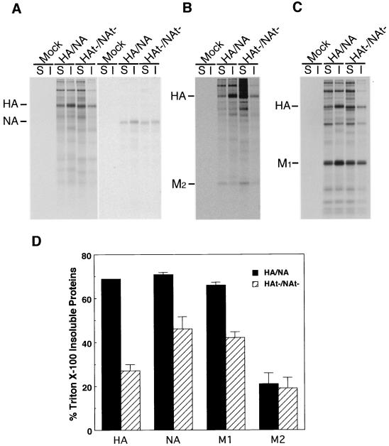
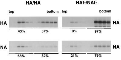
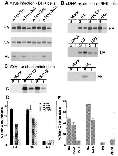
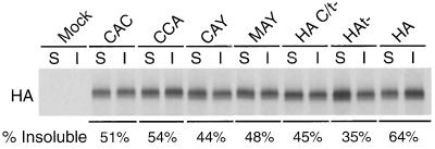
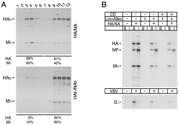
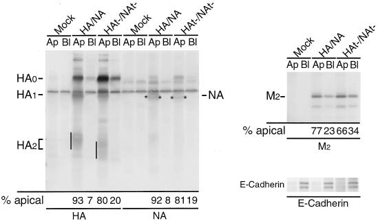
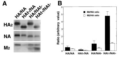
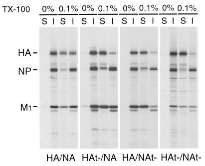
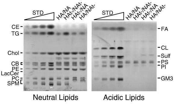
Influenza viruses encoding hemagglutinin (HA) and neuraminidase (NA) glycoproteins with deletions in one or both cytoplasmic tails (HAt- or NAt-) have a reduced association with detergent-insoluble glycolipids (DIGs). Mutations which eliminated various combinations of the three palmitoylation sites in HA exhibited reduced amounts of DIG-associated HA in virus-infected cells. The influenza virus matrix (M(1)) protein was also found to be associated with DIGs, but this association was decreased in cells infected with HAt- or NAt- virus. Regardless of the amount of DIG-associated protein, the HA and NA glycoproteins were targeted primarily to the apical surface of virus-infected, polarized cells. The uncoupling of DIG association and apical transport was augmented by the observation that the influenza A virus M(2) protein as well as the influenza C virus HA-esterase-fusion glycoprotein were not associated with DIGs but were apically targeted. The reduced DIG association of HAt- and NAt- is an intrinsic property of the glycoproteins, as similar reductions in DIG association were observed when the proteins were expressed from cDNA. Examination of purified virions indicated reduced amounts of DIG-associated lipids in the envelope of HAt- and NAt- viruses. The data indicate that deletion of both the HA and NA cytoplasmic tails results in reduced DIG association and changes in both virus polypeptide and lipid composition.


Figures

FIG. 1
FIG. 1
TX-100 solubility of membrane proteins in HA/NA and HAt−/NAt− virus-infected MDCK cells. Influenza virus-infected MDCK cells were pulse-labeled with [35S]-Promix, cell surfaces were biotinylated, cells were extracted with 1% TX-100, and soluble (S) and insoluble (I) fractions were separated by centrifugation. Proteins were immunoprecipitated, biotinylated proteins were recovered with streptavidin-Sepharose beads, and polypeptides were analyzed by SDS-PAGE. (A) Biotinylated HA and NA; (B) biotinylated HA and M2; (C) total HA and M1 immunoprecipitated using anti-A/Udorn/72 influenza virus serum; (D) quantitation of TX-100-insoluble HA, NA, M1, and M2 proteins (data averaged from two independent experiments).
FIG. 2
FIG. 2
Reduced DIGs association of HAt− and NAt− in virus-infected MDCK cells. HA/NA or HAt−/NAt− virus-infected MDCK cells were pulse-labeled with [35S]-Promix, surface biotinylated, and extracted with TX-100. The lysate was then loaded at the bottom of a flotation sucrose density gradient and subjected to equilibrium centrifugation. The gradient was fractionated from the top, fractions were immunoprecipitated, biotinylated HA or NA was recovered by using streptavidin-Sepharose beads, and biotinylated polypeptides were analyzed by SDS-PAGE. The percentages of DIG-associated (top five fractions) and non-DIG-associated (bottom five fractions) proteins are indicated beneath the gels.
FIG. 3
FIG. 3
TX-100 solubility of membrane proteins in BHK cells infected with HA/NA, HAt−/NA, HA/NAt−, and HAt−/NAt−. Virus-infected or plasmid-transfected BHK cells were pulse-labeled with [35S]-Promix, cell surfaces were biotinylated and extracted with 1% TX-100, and soluble (S) and insoluble (I) fractions were separated by centrifugation. Proteins were immunoprecipitated, surface biotinylated proteins were recovered with streptavidin-Sepharose beads, and polypeptides were analyzed by SDS-PAGE. (A) Surface HA, NA, and M2 in virus-infected BHK cells; (B) surface HA, NA, and M2 in plasmid-transfected BHK cells; (C) surface G protein from VSV-infected (VSV Gi) and VSV G cDNA-transfected (VSV Gt) BHK cells; (D) quantification of TX-100-insoluble HA, NA, and M2 proteins in virus-infected BHK cells; (E) quantification of TX-100-insoluble HA, NA, M2, and G proteins expressed from cDNAs. Panels D and E represent the average data from two independent experiments.
FIG. 4
FIG. 4
Palmitoylation and the cytoplasmic tail of HA contribute to its resistance to TX-100 extraction. (A) MDCK cells were infected with various influenza A viruses harboring the indicated mutations in HA. TX-100-soluble and -insoluble surface HA proteins were analyzed as described in the legend to Fig. 1. (B) Quantification of TX-100-insoluble HA proteins (data averaged from two independent experiments).
FIG. 5
FIG. 5
The TX-100 insolubility of M1 protein in influenza virus-infected cells. (A) HA/NA and HAt−/NAt− virus-infected MDCK cells were subjected to TX-100 extraction and flotation gradient treatment as described in the legend to Fig. 2. Fractions were taken from the top and immunoprecipitated with goat sera with specificity for HA and M1, and polypeptides were analyzed by SDS-PAGE. (B) BHK cells were untreated or treated with lovastatin-mevalonate (Lov+Mev) and, where indicated, with methyl-β-cyclodextrin (CD). Cell were then infected with HA/NA influenza virus or VSV, pulse-labeled with [35S]-Promix, and extracted with 1% TX-100 at 4°C. Soluble (S) and insoluble (I) fractions were separated by centrifugation and immunoprecipitated, and polypeptides were analyzed by SDS-PAGE.
FIG. 6
FIG. 6
Deletion of the cytoplasmic tails of HA and NA does not affect their transport to the apical cell surface. Polarized MDCK cells grown in Transwell inserts were infected with the indicated virus, and cell surfaces were biotinylated from either the apical (Ap) or the basolateral (Bl) side at 5 h p.i. The proteins of interest were immunoprecipitated, separated by electrophoresis, blotted to PVDF membranes, and detected by ECF blot assay. Quantification of the data is shown beneath the blots. The partial cleavage of HA0 reflects the A/WSN/33 NA-mediated cleavage of A/Udorn/72 HA by residual plasminogen in the medium (14). No exogenous trypsin was added. Asterisks indicates NA polypeptide.
FIG. 7
FIG. 7
The influenza C virus HEF protein is TX-100 soluble and transported to the apical surface. Influenza C/Ann Arbor/1/50 virus-infected MDCK cells (A) or HEF cDNA-transfected HeLa-T4 cells (B) were pulse-labeled with [35S]-Promix and after the indicated chase times incubated with TPCK-trypsin (15 μg/ml) for 10 min at 37°C to cleave cell surface-expressed HEF0 to HEF1 and HEF2. Cells were extracted with 1% TX-100, soluble (S) and insoluble (I) fractions were separated by centrifugation, HEF was immunoprecipitated, and polypeptides were analyzed by SDS-PAGE. (C) The apical (Ap) and basolateral (Bl) transport of HEF in infected MDCK cells was analyzed as described in the legend to Fig. 6.
FIG. 8
FIG. 8
Incorporation of M2 into HA and NA cytoplasmic tail-altered influenza viruses. Virions were purified on sucrose gradients, N-linked carbohydrate chains were digested with peptidyl N-glycosidase F, and polypeptides were separated by SDS-PAGE and blotted to PVDF membranes. HA, NA, and M2 proteins were detected with specific antibodies using an ECF Western blot assay (Pharmacia Amersham Biotech) (A). Quantification of the data was performed using ImageQuant software (Molecular Dynamics), and M2/NA and M2/HA ratios were plotted (B). The faster-migrating species of M2 is a proteolytic product of M2 and was included in the quantification.
FIG. 9
FIG. 9
TX-100 solubility of HA and M1 protein in HA and NA cytoplasmic tail-altered influenza viruses. Purified MDCK cell-grown 35S-labeled influenza viruses as indicated were extracted with 0.1% TX-100 for 30 min at 4°C. Soluble (S) and insoluble (I) fractions were separated by centrifugation, and polypeptides were analyzed by SDS-PAGE.
FIG. 10
FIG. 10
Lipid composition analysis of HA and NA cytoplasmic tail-altered influenza viruses. Lipids were extracted from purified HA/NA, HAt−/NA, HA/NAt−, and HAt−/NAt− virions grown in embryonated eggs as described in Materials and Methods. Lipids were analyzed by HPTLC. Wedges: 1, 3, 5, and 10 μg, in the four lanes, respectively, of each standard lipid. The experiment was done in duplicate, and representative TLC plates are shown. CE, cholesteryl ester; TG, triglyceride; Chol, cholesterol; CB, cerebroside; PE, phosphotidylethanolamine; LacCer, lactosyl ceramide; PC, phosphotidylcholine; SPM, sphingomyelin; FA, fatty acids; CL, cardiolipin; Sulf, sulfatides; PS, phosphotidylserine; PI, phosphotidylinositol.

References

    1. Avalos R T, Yu Z, Nayak D P. Association of influenza virus NP and M1 proteins with cellular cytoskeletal elements in influenza virus-infected cells. J Virol. 1997;71:2947–2958. - PMC - PubMed
    1. Brown D A, Crise B, Rose J K. Mechanism of membrane anchoring affects polarized expression of two proteins in MDCK cells. Science. 1989;245:1499–1501. - PubMed
    1. Brown D A, London E. Functions of lipid rafts in biological membranes. Annu Rev Cell Dev Biol. 1998;14:111–136. - PubMed
    1. Brown D A, Rose J K. Sorting of GPI-anchored proteins to glycolipid-enriched membrane subdomains during transport to the apical cell surface. Cell. 1992;68:533–544. - PubMed
    1. Burnet F M. Growth of influenza virus in the allantoic cavity of the chick embryo. Aust J Exp Biol Med Sci. 1941;19:291–295.

Publication types

MeSH terms

LinkOut - more resources