. 2000 May;74(10):4765-75.
doi: 10.1128/jvi.74.10.4765-4775.2000.

Complement depletion facilitates the infection of multiple brain tumors by an intravascular, replication-conditional herpes simplex virus mutant

Affiliations

Complement depletion facilitates the infection of multiple brain tumors by an intravascular, replication-conditional herpes simplex virus mutant

K Ikeda et al. J Virol. 2000 May.

Abstract

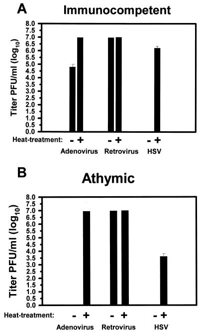
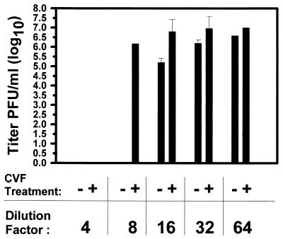
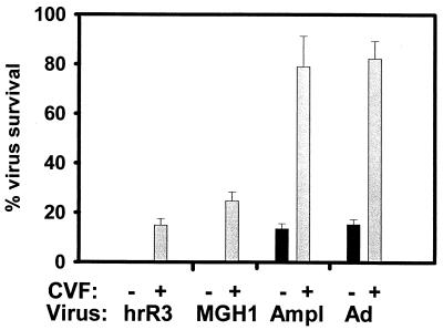
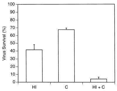
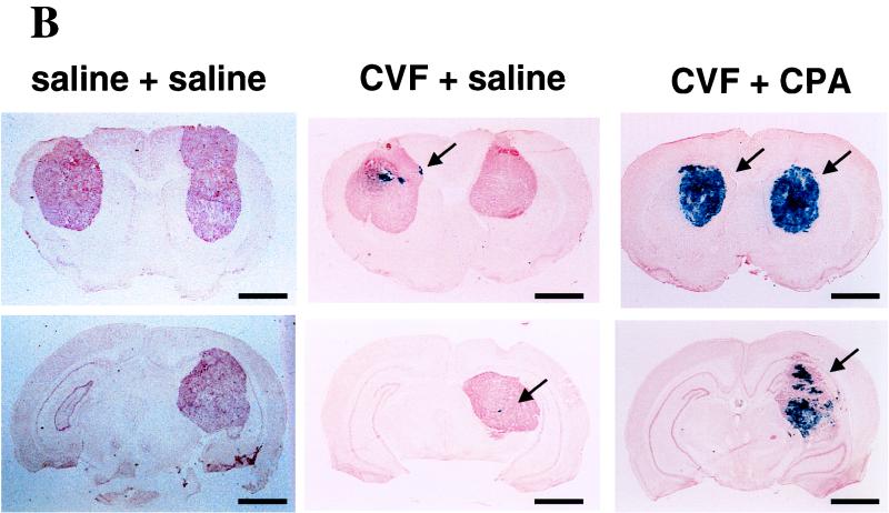
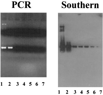
Intravascular routes of administration can provide a means to target gene- and virus-based therapies to multiple tumor foci located within an organ, such as the brain. However, we demonstrate here that rodent plasma inhibits cell transduction by replication-conditional (oncolytic) herpes simplex viruses (HSV), replication-defective HSV, and adenovirus vectors. In vitro depletion of complement with mild heat treatment or in vivo depletion by treatment of athymic rats with cobra venom factor (CVF) partially reverses this effect. Without CVF, inhibition of cell infection by HSV is observed at plasma dilution as high as 1:32, while plasma from CVF-treated animals displays anti-HSV activity at lower dilutions (1:8). When applied to the therapy of intracerebral brain tumors, in vivo complement depletion facilitates the initial infection (assayed at the 2-day time point) by an intra-arterial replication-conditional HSV of tumor cells, located within three separate and distinct human glioma masses. However, at the 4-day time point, no propagation of HSV from initially infected tumor cells could be observed. Previously, we have shown that the immunosuppressive agent, cyclophosphamide (CPA), facilitates the in vivo propagation of an oncolytic HSV, delivered intravascularly, within infected multiple intracerebral masses, by inhibition of both innate and elicited anti-HSV neutralizing antibody response (K. Ikeda et al., Nat. Med. 5:881-889, 1999). In this study, we thus show that the addition of CPA to the CVF treatment results in a significant increase in viral propagation within infected tumors, measured at the 4-day time period. The concerted action of CVF and CPA significantly increases the life span of athymic rodents harboring three separate and large glioma xenografts after treatment with intravascular, oncolytic HSV. Southern analysis of viral genomes analyzed by PCR reveals the presence of the oncolytic virus in the brains, livers, spleens, kidneys, and intestine of treated animals, although none of these tissues displays evidence of HSV-mediated gene expression. In light of clinical trials of oncolytic HSV for malignant brain tumors, these findings suggest that antitumor efficacy may be limited by the host innate and elicited humoral responses.


Figures

FIG. 1
FIG. 1
The in vitro effect of control and heat-treated (complement-depleted) rodent plasma on virus vector infectivity of cells. Plasma was prepared from immunocompetent (A) and athymic (B) rats as described in Materials and Methods and either exposed to a temperature of 56°C for 30 min or not before incubation with an adenovirus, retrovirus, or HSV vector expressing the lacZ cDNA for 1.5 h. The viruses were then added onto Vero cells and, 16 h later, the number of lacZ cDNA-expressing cells was recorded. Values represent the mean titer of virus from triplicate dishes. In pilot experiments, virus preincubation with HBSS or with HBSS-bovine serum albumin (10 mg/ml) did not result in significant reductions of virus titers (data not shown). Error bars represent the standard error of the mean. If not shown, then the error bars were too small to graph.
FIG. 2
FIG. 2
Plasma anti-HSV activity and partial reversal by CVF. Plasma was prepared from athymic rats 48 h after intraperitoneal administration of CVF (or carrier) as detailed in Materials and Methods. After serial dilutions, it was mixed with oncolytic HSV before it was added onto Vero cells. Values represent the mean titer of virus from triplicate dishes. Controls, consisting of virus preincubated with HBSS for the same times before addition onto plates, did not show a reduction in mean titer (data not shown). Error bars represent the standard error of the mean. If not shown, then the error bars were too small to graph.
FIG. 3
FIG. 3
Effects of CVF-treated and control plasma on three HSV vectors and adenovirus. Plasma was prepared from athymic rats 2 days after treatment with CVF or carrier. It was then diluted 1:8 before being mixed with hrR3, an ICP6-defective HSV derived from KOS strain; MGH1, an ICP6- and ICP34.5-defective HSV derived from F strain; Amplicon (Ampl), a replication-defective HSV amplicon derived from strain 17; or an adenoviral (Ad) vector. The percent virus survival denotes the percentage of lacZ-expressing Vero cells, as enumerated 16 h after infection, compared to control dishes. Controls consisted of virus preincubated with HBSS for the same times before addition onto plates. The bars represent the average from triplicate dishes, and error bars represent the standard error of the mean.
FIG. 4
FIG. 4
Rat complement inactivates oncolytic HSV. The hrR3 mutant virus was preincubated with HBSS, purified rat complement (C; 1 mg/ml in HBSS), heat-inactivated rat plasma (HI; diluted 1:4), or rat complement re-added to heat-inactivated plasma (C + HI) for 1.5 h before adding it onto Vero cells in culture. The percent virus survival denotes the percentage of lacZ-expressing Vero cells (as enumerated 16 h after infection) compared to control dishes. Controls consisted of virus preincubated with HBSS for the same times before addition onto plates. In pilot experiments, virus preincubation with HBSS or with HBSS-bovine serum albumin (10 mg/ml) did not result in significant differences (data not shown). The bars represent the average from duplicate dishes, and the error bars represent the standard error of the mean.
FIG. 5
FIG. 5
Histologic sections of brains with three neoplasms at 2 (A), 4 (B), and 8 (C) days after intravascular treatment with hrR3 in the presence or absence of CVF and CPA. Human U87dEGFR glioma cells were implanted into three separate intracerebral locations (right and left frontal lobes and right thalamus). Animals were treated with CVF (60 U/kg) or saline 6 days later and with another dose of CVF (20 U/kg) or saline 7 days later. At this time point, animals were treated with intravascular hrR3 and RMP7. Some animals also received an intraperitoneal injection of CPA (100 mg/kg) 2 days after intravascular treatment with hrR3. Animals were sacrificed 2, 4, and 8 days after virus administration, and their brains were harvested, sectioned, and stained for lacZ cDNA expression. The anatomic extent of tumor transduction was measured for each of the three neoplasms and tabulated in Table 3. Photomicrographs of sections showing brains with tumors stained for lacZ cDNA expression reveal transgene expression in a “plaque”-like configuration within tumors from CVF treated-animals at 2 days after administration of hrR3 (arrow in panel A). Four days later, the anatomic area of the lacZ-expressing plaques has not increased in the CVF-treated animals. However, the addition of CPA results in a significant augmentation of lacZ-expressing cells within the tumors (arrows in panel B). By 8 days, the oncolytic action of the virus has resulted in tumor involution (arrows in panel C). Bars, 4 mm.
FIG. 5
FIG. 5
Histologic sections of brains with three neoplasms at 2 (A), 4 (B), and 8 (C) days after intravascular treatment with hrR3 in the presence or absence of CVF and CPA. Human U87dEGFR glioma cells were implanted into three separate intracerebral locations (right and left frontal lobes and right thalamus). Animals were treated with CVF (60 U/kg) or saline 6 days later and with another dose of CVF (20 U/kg) or saline 7 days later. At this time point, animals were treated with intravascular hrR3 and RMP7. Some animals also received an intraperitoneal injection of CPA (100 mg/kg) 2 days after intravascular treatment with hrR3. Animals were sacrificed 2, 4, and 8 days after virus administration, and their brains were harvested, sectioned, and stained for lacZ cDNA expression. The anatomic extent of tumor transduction was measured for each of the three neoplasms and tabulated in Table 3. Photomicrographs of sections showing brains with tumors stained for lacZ cDNA expression reveal transgene expression in a “plaque”-like configuration within tumors from CVF treated-animals at 2 days after administration of hrR3 (arrow in panel A). Four days later, the anatomic area of the lacZ-expressing plaques has not increased in the CVF-treated animals. However, the addition of CPA results in a significant augmentation of lacZ-expressing cells within the tumors (arrows in panel B). By 8 days, the oncolytic action of the virus has resulted in tumor involution (arrows in panel C). Bars, 4 mm.
FIG. 6
FIG. 6
Kaplan-Meier survival analyses of athymic rats harboring three separate human glioma xenografts. (A) Survival of rats injected with hrR3. (B) Survival of rats mock injected. Ten animals were treated in each group. Arrows indicate the time point of catheterization. In the hrR3-treated group, the differences in survival between animals treated with CVF+CPA versus CVF alone or CPA alone were both statistically significant (P < 0.001, Wilcoxon signed rank test). The addition of RMP7 to the hrR3+CVF+CPA treatment produced a slight increase in survival but was not statistically significant (P = 0.2). All treatments with hrR3 were significant compared to treatments without virus (P < 0.001).
FIG. 7
FIG. 7
Analysis of viral genomes in athymic rat tissues after intra-arterial administration of hrR3 in the presence of CVF and CPA. Genomic DNA was prepared from tissues of brain tumor (lane 2), brain surrounding the tumor (lane 3), lungs (lane 4), livers (lane 5), spleens (lane 6), and kidneys (lane 7) from athymic rats and was then analyzed by PCR using primers specific for hrR3. Lane 1 represents a positive control consisting of plasmid DNA containing the expected ICP6::lacZ cDNA sequence. The 5′ primer hybridizes to the 5′ region of the HSV ICP6 genome, and the 3′ primer hybridizes to the 5′ region of the inserted lacZ cDNA. After agarose gel electrophoresis and ethidium bromide staining (shown in the panel on the left), Southern analysis of the PCR products was performed using a lacZ cDNA probe that should hybridize to the amplified PCR fragments. The size of the PCR product is approximately 1,000 bp.

References

    1. Aghi M, Chou T C, Suling K, Breakefield X O, Chiocca E A. Multimodal cancer treatment mediated by a replicating oncolytic virus that delivers the oxazaphosphorine/rat cytochrome P450 2B1 and ganciclovir/herpes simplex virus thymidine kinase gene therapies. Cancer Res. 1999;59:3861–3865. - PubMed
    1. Anderson S C, Johnson D E, Harris M P, Engler H, Hancock W, Huang W M, Wills K N, Gregory R J, Sutjipto S, Wen S F, Lofgren S, Shepard H M, Maneval D C. p53 gene therapy in a rat model of hepatocellular carcinoma: intra-arterial delivery of a recombinant adenovirus. Clin Cancer Res. 1998;4:1649–1659. - PubMed
    1. Ash R J. Butyrate-induced reversal of herpes simplex virus restriction in neuroblastoma cells. Virology. 1986;155:584–592. - PubMed
    1. Ballow M, Cochrane C G. Two anticomplementary factors in cobra venom: hemolysis of guinea pig erythrocytes by one of them. J Immunol. 1969;103:944–952. - PubMed
    1. Barnard R O, Geddes J F. The incidence of multifocal cerebral gliomas. A histologic study of large hemisphere sections. Cancer. 1987;60:1519–1531. - PubMed

Publication types

MeSH terms