. 2000 May;74(10):4831-8.
doi: 10.1128/jvi.74.10.4831-4838.2000.

Rescue of mumps virus from cDNA

Affiliations

Rescue of mumps virus from cDNA

D K Clarke et al. J Virol. 2000 May.

Abstract

A complete DNA copy of the genome of a Jeryl Lynn strain of mumps virus (15,384 nucleotides) was assembled from cDNA fragments such that an exact antigenome RNA could be generated following transcription by T7 RNA polymerase and cleavage by hepatitis delta virus ribozyme. The plasmid containing the genome sequence, together with support plasmids which express mumps virus NP, P, and L proteins under control of the T7 RNA polymerase promoter, were transfected into A549 cells previously infected with recombinant vaccinia virus (MVA-T7) that expressed T7 RNA polymerase. Rescue of infectious virus from the genome cDNA was demonstrated by amplification of mumps virus from transfected-cell cultures and by subsequent consensus sequencing of reverse transcription-PCR products generated from infected-cell RNA to verify the presence of specific nucleotide tags introduced into the genome cDNA clone. The only coding change (position 8502, A to G) in the cDNA clone relative to the consensus sequence of the Jeryl Lynn plaque isolate from which it was derived, resulting in a lysine-to-arginine substitution at amino acid 22 of the L protein, did not prevent rescue of mumps virus, even though an amino acid alignment for the L proteins of paramyxoviruses indicates that lysine is highly conserved at that position. This system may provide the basis of a safe and effective virus vector for the in vivo expression of immunologically and biologically active proteins, peptides, and RNAs.


Figures

FIG. 1
FIG. 1
Diagram (not to scale) showing the organization of the MUVCAT minireplicon DNA construct and T7 RNA polymerase-transcribed minireplicon antisense RNA genome. Key restriction endonuclease sites used in the assembly of the DNA construct are shown. The T7 RNA polymerase promoter sequence was designed to start transcription with the exact MUV 5′-terminal nucleotide, and an HDV ribozyme (Rib.) sequence was positioned to generate the precise MUV 3′-terminal nucleotide in minireplicon RNA transcripts. Duplicate T7 RNA polymerase termination signals were included after the HDV ribozyme sequence. The CAT ORF replaces all of the coding and intercistronic sequence of the MUV genome; the remaining essential MUV-specific sequence comprises the 3′ MUV leader (55 nt) with adjacent 90-nt NP gene untranslated region (UTR) and the 5′ MUV trailer (24 nt) adjacent to the 137-nt L gene untranslated region.
FIG. 2
FIG. 2
Schematic representation (not to scale) of the MUV full-length genome cDNA construct showing the genetic organization of the MUV genome including the nontranscribed leader (Le) and trailer (Tr) and the proteins expressed from each gene. The subgenomic cDNA fragments and restriction endonuclease sites used in the assembly process are delineated by the horizontal solid lines and the vertical dotted lines, respectively. The T7 RNA polymerase promoter (T7-P) and the HDV ribozyme (Rib) sequence were positioned to initiate transcription with the exact 5′-terminal nucleotide and generate the precise 3′-terminal nucleotide of the MUV antisense genome, respectively. Tandem T7 RNA polymerase termination sequences were placed adjacent to the HDV ribozyme.
FIG. 3
FIG. 3
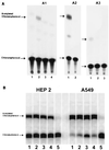
(A) Thin-layer chromatograms showing CAT activity in 293 cells following infection with MUV and transfection with RNA transcribed in vitro from pMUVCAT. Panels A1, A2, and A3 show the results from three separate rescue experiments. (A1) Lane 1, CAT activity in MUV-infected cells transfected without in vitro-transcribed pMUVCAT RNA; lane 2, CAT activity in extracts of MUV-infected cells transfected with RNA transcribed in vitro from pMUVCAT; lane 3, CAT activity in MUV-infected cells transfected with RNA transcribed in vitro from pMUVCAT-GG; lane 4, CAT activity in uninfected cells transfected with RNA transcribed in vitro from pMUVCAT. Each CAT assay was carried out at 37°C for 3 to 4 h with 20% of the extract from ∼106 transfected cells. (A2) Lane 1, MUV-infected cells transfected with RNA transcribed in vitro from pMUVCAT; lane 2, uninfected cells transfected with RNA transcribed in vitro from pMUVCAT. Each CAT assay was carried out at 37°C for 5 h using 50% of the extract from ∼106 transfected cells. (A3) Lane 1, MUV-infected cells transfected with RNA transcribed in vitro from pMUVCAT; lane 2, MUV-infected cells transfected without in vitro-transcribed pMUVCAT RNA; lane 3, uninfected cells transfected with in vitro-transcribed RNA from pMUVCAT. Each CAT assay shown in panel A3 was carried out at 37°C for 4 h using 50% of the extract from ∼106 transfected cells. (B) Thin-layer chromatograms showing CAT activity in extracts of MVA-T7-infected HEp-2 and A549 cells following transfection with pMUVCAT and plasmids expressing MUV NP, P, and L proteins. The level of pMUVNP expression plasmid was titrated in both cell lines. Lanes 1 to 4, CAT activity following transfection with mixtures containing 200 ng of pMUVCAT, 50 ng of pMUVP, and 200 ng of pMUVL each, and 300, 450, 600, and 750 ng of pMUVNP, respectively; lane 5, CAT activity produced when pMUVL was omitted from the transfection mixture. Each CAT assay was performed at 37°C for 3 h using 20% of the cell extract from each well of transfected cells (∼106 cells/well of a six-well dish).
FIG. 4
FIG. 4
(A) Photographs showing rMUV-induced syncytia on Vero cell monolayers. Supernatants from MVA-T7-infected A549 cells transfected with pMUVFL, pMUVNP, pMUVP, and pMUVL were passaged onto A549 cells, incubated at 37°C for 4 days, then transferred onto Vero cell monolayers, and incubated for 3 more days at 37°C prior to photography (A1). (A2) Representative portion of Vero cell monolayer following transfer of supernatant from transfected A549 cells as described for panel A1 except that pMUVL was omitted from the transfection mixture; (A3) syncytia produced on Vero cells following infection with Jeryl Lynn vaccine virus. (B) Photographs showing rMUV-induced plaques on Vero cell monolayers stained by whole-cell ELISA. Supernatant from transfected cells was passed onto A549 indicator cells and incubated for 3 days at 37°C; supernatant from these cells was then passed onto Vero cell monolayers. One of the resulting syncytia was picked and used to infect fresh Vero cell monolayers. Virus-induced plaques were then stained by whole-cell ELISA 4 days postinfection (B1) and compared to plaques induced by Jeryl Lynn vaccine virus (B3). Panel B2 shows Vero cells infected with cell supernatants as for cells in panel B1 except that the L expression plasmid was omitted from the starting transfection mixture.
FIG. 5
FIG. 5
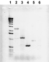
Gel analysis of RT-PCR products used to identify rMUV. Total RNA was prepared from Vero cell monolayers infected with P2 rMUV virus from transfected cells. RT-PCRs were set up to generate cDNA products spanning the three separate nucleotide tag sites present only in pMUVFL and rMUV. Lane 1, marker 1-kb ladder (Gibco/BRL); lanes 2 to 4, RT-PCR products spanning nucleotide tag positions 6081, 8502, and 11731, respectively. To demonstrate the absence of contaminating plasmid DNA, a reaction identical to that used for generation of the cDNA shown in lane 4 was performed without RT; the product(s) of this reaction is shown in lane 5. To demonstrate that no rMUV could be recovered when pMUVL was omitted from transfection mixtures, RT-PCR identical to that used to generate the cDNA products shown in lane 4 was set up using Vero cell RNA derived from transfections carried out without pMUVL; products from this reaction are shown in lane 6.
FIG. 6
FIG. 6
Electropherograms showing nucleotide sequence across identifying tag sites in rMUV. RT-PCR products (Fig. 5) were sequenced across each of the three tag sites. The nucleotide sequence at each tag site obtained for rMUV is compared with consensus sequence for the plaque isolate of MUV used to derive pMUVFL.

References

    1. Afzal M A, Pickford A R, Forsey T, Heath A B, Minor P D. The Jeryl Lynn vaccine strain of mumps virus is a mixture of two distinct isolates. J Gen Virol. 1993;74:917–920. - PubMed
    1. Baron M D, Barrett T. Rescue of rinderpest virus from cloned cDNA. J Virol. 1997;71:1265–1271. - PMC - PubMed
    1. Bucholz U J, Finke S, Conzelmann K-K. Generation of bovine respiratory syncytial virus (BRSV) from cDNA: BRSV NS2 is not essential for virus replication in tissue culture, and the human RSV leader region acts as a functional BRSV genome promoter. J Virol. 1999;73:251–259. - PMC - PubMed
    1. Calain P, Roux L. The rule of six, a basic feature for efficient replication of Sendai virus defective interfering RNA. J Virol. 1993;67:4822–4830. - PMC - PubMed
    1. Collins P L, Mink M A, Stec D S. Rescue of synthetic analogs of respiratory syncytial virus genomic RNA and effect of truncations and mutations on the expression of a foreign reporter gene. Proc Natl Acad Sci USA. 1991;88:9663–9667. - PMC - PubMed

Associated data