. 2000 May 1;20(9):3104-14.
doi: 10.1523/JNEUROSCI.20-09-03104.2000.

Involvement of retinoblastoma family members and E2F/DP complexes in the death of neurons evoked by DNA damage

Affiliations

Involvement of retinoblastoma family members and E2F/DP complexes in the death of neurons evoked by DNA damage

D S Park et al. J Neurosci. .

Abstract

Neuronal death evoked by DNA damage requires cyclin-dependent kinase 4 (Cdk4) and 6 activity and is accompanied by elevation of cyclin D1-associated kinase activity. Because Cdk4/6 phosphorylates retinoblastoma protein (pRb) family members that then modulate the transcriptional activity of E2F/DP1 complexes, we examined the involvement of these components in DNA damage-evoked neuronal death. Camptothecin induced rapid pRb and p107 phosphorylation at a Cdk4/6 phosphorylation site followed by selective loss of Rb and p107. The CDK inhibitor flavopiridol suppressed pRb and p107 phosphorylation and loss, implicating CDK activity in these events. Moreover, the loss of pRb and p107 appeared to be mediated by caspases because it was blocked by general caspase inhibitors. The role of phosphorylation and pRb and p107 loss in the death pathway was indicated by observations that virally mediated expression of pRb mutated at sites of phosphorylation, including the Cdk4/6 site, inhibited death. Finally, expression of dominant-negative versions of DP1, known to compromise E2F transcriptional activity, protects cortical neurons from death induced by camptothecin and sympathetic neurons from death evoked by UV treatment. Taken together, these results implicate the CDK-pRb/E2F/DP pathway as a required element in the neuronal death evoked by DNA damage.


Figures
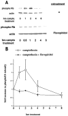
Fig. 1.
Fig. 1.
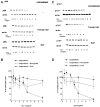
pRb/p107 is phosphorylated during the death of cortical neurons evoked by camptothecin. A, Top, Western immunoblots (probed with anti-phospho-Rb antibody) of whole-cell lysates of cortical neurons after various periods of treatment with camptothecin (10 μm). Bottom, Cotreatment with flavopiridol (1 μm) is indicated.campto, Camptothecin. B, Densitometric analyses of Western immunoblots of cortical neurons treated with camptothecin alone or cotreated with flavopiridol and probed with anti-phospho-Rb antibody. All data points are the mean ± SEM of data from three separate experiments and are expressed relative to the initial amount of phosphorylated pRb/p107 at time 0.
Fig. 2.
Fig. 2.
pRb and p107 levels decrease during the death of cortical neurons evoked by camptothecin. A, Top, C, Top, Representative Western immunoblots of whole-cell extracts of cortical neurons treated with camptothecin for the indicated times and probed with antibodies for pRb (A) and p107 (C). Middle,Bottom, Cotreatment with flavopiridol (1 μm; middle) and BAF (100 μm;bottom) is indicated. Blots were stripped and reprobed for β-actin as a loading control. B, D, Densitometric analyses of Western immunoblots of cortical neurons treated with camptothecin alone or cotreated with flavopiridol or BAF and probed for pRb (B) or p107 (D). All data points are the mean ± SEM of data from three separate experiments and are expressed relative to the initial amount of protein at time 0.
Fig. 3.
Fig. 3.
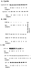
Levels of cyclins, CDKs, and CKIs do not increase during the death of cortical neurons evoked by camptothecin.A—C, Western immunoblot analyses of whole-cell extracts of cortical neurons treated with camptothecin for the indicated times and probed with the following antibodies: cyclin D1 and cyclin E (A); Cdk2, Cdk4, and Cdk6 (B); and p16, p21, and p27 (C). For Cdk2 blots, an equal amount (25 μg) of rat fibroblast cell extract was used as a positive control.
Fig. 4.
Fig. 4.
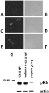
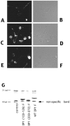
Immunofluorescence (A–F) and Western immunoblot (G) analyses of ΔK11 Rb expression. A—F, Immunofluorescence (A, C, E) staining with an antibody directed against pRb or corresponding light micrographs (B, D, F) of cortical neurons in culture infected with ΔK11 Rb-expressing virus (C–F) or control (A, B). Neurons were fixed and stained with anti-pRb antibody 24 hr after infection (A–D) and after 12 hr of camptothecin treatment (E, F). G, Western immunoblot analysis of whole-cell extracts of cortical neurons infected with adenovirus expressing ΔK11 Rb (no treatment or 10 hr camptothecin treatment) or LacZ as indicated. The blots were analyzed using an anti-pRb antibody, stripped, and reprobed with anti-β-actin.
Fig. 5.
Fig. 5.
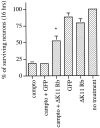
Expression of ΔK11 Rb suppresses the death of cortical neurons evoked by camptothecin. Neuronal cultures were uninfected or infected for 24 hr with adenovirus expressing ΔK11 Rb or GFP before camptothecin treatment for 16 hr. Each point is the mean ± SEM of combined data from three independent experiments (each independent experiment performed in triplicate) and is expressed relative to the number of neurons present at the initial time of camptothecin treatment. * indicates significance (p < 0.02, Student's ttest) compared with GFP-infected cultures treated with camptothecin.
Fig. 6.
Fig. 6.
A—F, Immunofluorescence (A, C, E) or corresponding phase (B, D, F) images of neurons infected with control virus (A, B) or with virus encoding (Δ103–126) DN DP1F (C–F). Neurons were fixed and stained with anti-FLAG antibody 24 hr after infection (A–D) and after 12 hr of camptothecin treatment (E, F). G, Western immunoblot analyses of whole-cell extracts of cortical neurons infected with Sindbis viruses expressing WT or the indicated mutant FLAG-tagged DP1 constructs. control indicates samples in which the neurons were infected with a control virus. Theblots were analyzed using an anti-FLAG antibody. A nonspecific immunoreactive band is marked as indicated. Neurons were lysed 24 hr after infection with the indicated viruses.
Fig. 7.
Fig. 7.
Expression of DN DP1 suppresses the death of cultured cortical neurons treated with camptothecin. Control viruses for each vector were generated by removal of the start codon from the insert. F denotes that the protein has a FLAG epitope attached to the C terminal. Effects of expression of (Δ1–126) DN DP1, a dominant-negative mutant containing an N-terminal truncation from amino acids 1–126 (A), (Δ103–126) DN DP1F, a FLAG-tagged dominant-negative mutant containing a DNA-binding domain deletion of amino acids 103–126 (B), (Δ233–272) DP1F, a FLAG-tagged mutant containing the E2F-binding domain deletion of amino acids 233–272 (C), and WT DP1 and respective controls (D) on the survival of cortical neurons after camptothecin treatment for 1 d. A, Graph in which each point is the mean ± SEM of combined data from three independent experiments (each independent experiment performed in triplicate) and is expressed relative to the number of neurons present in each culture at the initial time of camptothecin (10 μm) treatment. B—D, Representative experiments in which each point is the mean ± SEM of three cultures from the same experimental trial. Similar results (B–D) were obtained in two independent experiments.
Fig. 8.
Fig. 8.
Phase-contrast micrographs of cortical neurons treated for 1 d with the following: no additives (A), camptothecin (10 μm;B), (Δ1–126) DN DP1 virus + camptothecin (C), and (Δ1–126) DN DP1 control virus + camptothecin (D).
Fig. 9.
Fig. 9.
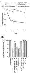
Expression of DN DP1 suppresses the death of cultured sympathetic neurons evoked by UV irradiation. Control viruses for each vector were generated by removal of the start codon from the insert. F denotes that the protein has a FLAG epitope attached to the C terminal. Effects of expression of (Δ1–126) DN DP1, a dominant-negative mutant containing an N-terminal truncation from amino acids 1–126 (A), and of (Δ103–126) DN DP1F, a FLAG-tagged dominant-negative mutant containing a DNA-binding domain deletion of amino acids 103–126, (Δ233–272) DP1F, a FLAG-tagged mutant containing the E2F-binding domain deletion of amino acids (233–272), and wild type (WT) DP1 (B) on the survival of sympathetic neurons after UV irradiation. A, Graph in which each point is the mean ± SEM of combined data from three independent experiments (each independent experiment performed in triplicate) and is expressed relative to the number of neurons present in each culture at the initial time of UV treatment. cont, Control.B, Representative experiment in which each point is the mean ± SEM of three cultures from the same experimental trial. Similar results were obtained in two independent experiments.
Fig. 10.
Fig. 10.
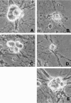
Phase-contrast micrographs of rat sympathetic neurons maintained in NGF-containing medium and exposed to and/or infected with the following: no treatment (A), UV (B), (Δ1–126) DN DP1 virus + UV (C), (Δ1–126) DN DP1 control virus + UV (D), or (Δ1–126) DN DP1 virus alone (E). The photos were taken 2 d after irradiation and/or infection.
Fig. 11.
Fig. 11.
Immunofluorescence (A–C) staining with an antibody directed against the FLAG epitope or corresponding light micrographs (D–F) of sympathetic neurons in culture infected with Sindbis virus expressing (Δ233–272) DP1F (A, D) or (Δ103–126) DN DP1F (B, E) or containing a nonexpressing control virus (C, F). Neurons were stained 2 d after infection.

Similar articles

Cited by

References

    1. Anderson AJ, Su JH, Cotman CW. DNA damage and apoptosis in Alzheimer's disease: colocalization with c-jun immunoreactivity, relationship to brain area, and effect of postmortem delay. J Neurosci. 1996;16:1710–1719. - PMC - PubMed
    1. Anderson CNG, Tolkovsky AM. A role for MAPK/ERK in sympathetic neuron survival: protection against a p53-dependent, JNK-independent induction of apoptosis by cytosine arabinoside. J Neurosci. 1999;19:664–673. - PMC - PubMed
    1. Bandara LR, Buck VM, Zamanian M, Johnston LH, La Thangue NB. Functional synergy between DP-1 and E2F-1 in the cell cycle-regulating transcription factor DRTF1/E2F. EMBO J. 1993;12:4317–4324. - PMC - PubMed
    1. Beijersbergen RL, Carlee L, Kerkhoven RM, Bernards R. Regulation of the retinoblastoma protein-related p107 by G1 cyclin complexes. Genes Dev. 1995;9:1340–1350. - PubMed
    1. Berry DE, Lu Y, Schimdt B, Falon PG, O'Connell C, Hu S-X, Xu H-J, Blanck G. Retinoblastoma protein inhibits IFN induced apoptosis. Oncogene. 1996;12:1809–1819. - PubMed

Publication types

MeSH terms

Substances