. 2000 May 1;20(9):3221-32.
doi: 10.1523/JNEUROSCI.20-09-03221.2000.

Brain-derived neurotrophic factor differentially regulates excitatory and inhibitory synaptic transmission in hippocampal cultures

Affiliations

Brain-derived neurotrophic factor differentially regulates excitatory and inhibitory synaptic transmission in hippocampal cultures

M M Bolton et al. J Neurosci. .

Abstract

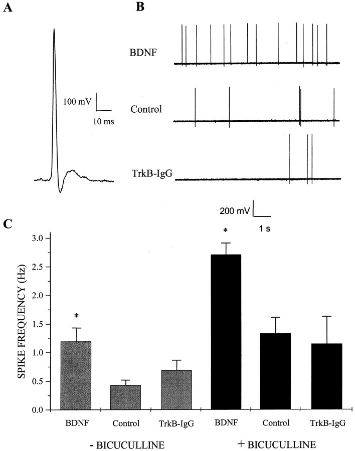
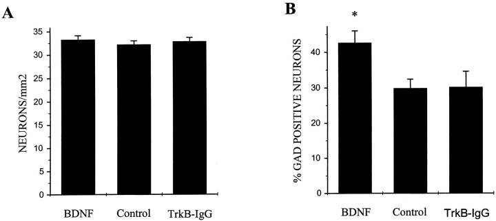
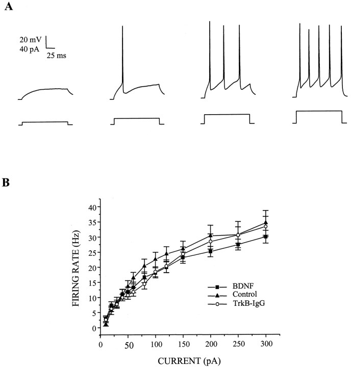
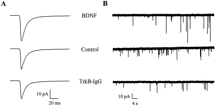
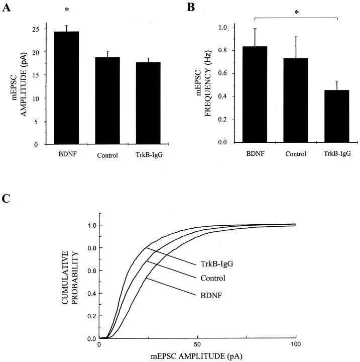
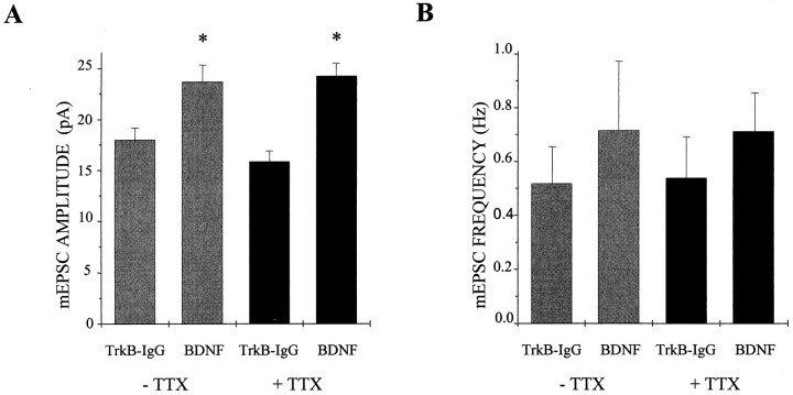
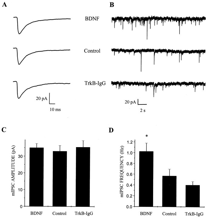
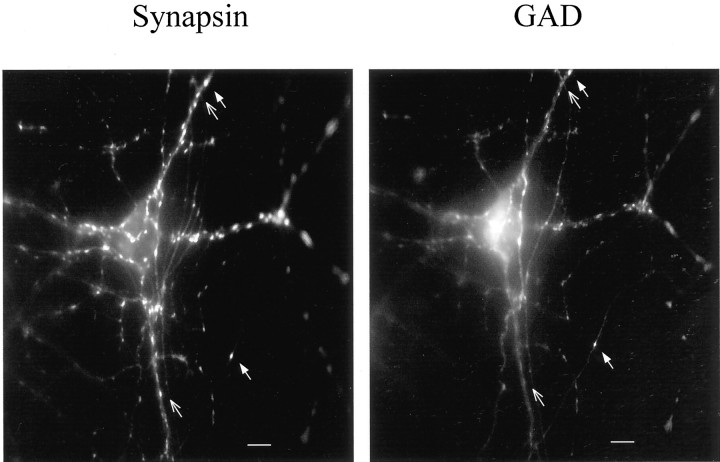
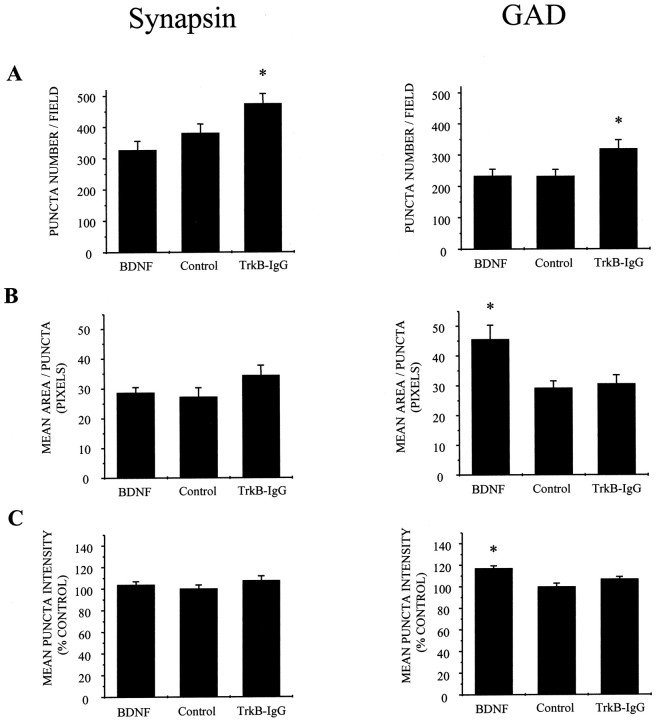
Brain-derived neurotrophic factor (BDNF) has been postulated to be a key signaling molecule in regulating synaptic strength and overall circuit activity. In this context, we have found that BDNF dramatically increases the frequency of spontaneously initiated action potentials in hippocampal neurons in dissociated culture. Using analysis of unitary synaptic transmission and immunocytochemical methods, we determined that chronic treatment with BDNF potentiates both excitatory and inhibitory transmission, but that it does so via different mechanisms. BDNF strengthens excitation primarily by augmenting the amplitude of AMPA receptor-mediated miniature EPSCs (mEPSCs) but enhances inhibition by increasing the frequency of mIPSC and increasing the size of GABAergic synaptic terminals. In contrast to observations in other systems, BDNF-mediated increases in AMPA-receptor mediated mEPSC amplitudes did not require activity, because blocking action potentials with tetrodotoxin for the entire duration of BDNF treatment had no effect on the magnitude of this enhancement. These forms of synaptic regulations appear to be a selective action of BDNF because intrinsic excitability, synapse number, and neuronal survival are not affected in these cultures. Thus, although BDNF induces a net increase in overall circuit activity, this results from potentiation of both excitatory and inhibitory synaptic drive through distinct and selective physiological mechanisms.


Figures

Fig. 1.
Fig. 1.
BDNF increases spontaneous action potential firing rate. A, Representative traceillustrating the waveform of an action potential recorded with an on-cell patch pipette; this configuration was used to measure action potential frequency in dissociated hippocampal cultures. Voltage traces were inverted about the vertical axis to conform to convention. Data were acquired at 5 kHz in voltage-follower recording mode and filtered at 2 kHz. B, Spontaneous action potential firing was increased in cultures treated with BDNF. Representative on-cell recordings shown at a compressed time base from cells treated for 4–7 d with 100 ng/ml BDNF (top), untreated (Control, middle), or treated with 5 μg/ml TrkB-IgG (bottom). Cultures were rinsed in recording saline several times to ensure that no BDNF or TrkB-IgG was present at the time of recording. C,Left, BDNF treatment increased the spontaneous firing rate of pyramidal neurons approximately threefold compared with untreated controls; means ± SEM are shown. *p< 0.009 indicates a significant difference by ANOVA between BDNF and either control or TrkB-IgG treatment groups;n = 33, 27, and 36 for BDNF, control, and TrkB-IgG groups, respectively. Right, Elevated action potential firing rates induced by BDNF persisted in disinhibited circuits. BDNF appeared to increase excitatory synaptic transmission directly because the increase in spontaneous firing rates of pyramidal neurons persisted after acute blockade of inhibitory transmission by bicuculline during the recording period. *p < 3 × 10⁻⁵ indicates a significant difference by ANOVA between BDNF and either control or TrkB-IgG treatment groups;n = 43, 36, and 12 for BDNF, control, and TrkB-IgG groups, respectively.
Fig. 2.
Fig. 2.
BDNF enhances the phenotypic differentiation of GABAergic neurons but has no effect on neuronal survival. Hippocampal cultures were treated for 5 d with either BDNF or TrkB-IgG or were left untreated; cultures were then double-labeled for the neuronal marker NSE and the GABAergic neuronal marker GAD. Numbers of NSE-positive and GAD-positive neurons were counted independently; all counts were done blind. A, BDNF treatment did not affect neuronal survival. Means ± SEM are shown; n = 9 dishes per condition with 20 fields counted and averaged per dish; p > 0.35 for any pairwise comparison by ANOVA. B, BDNF increased the percentage of GAD-positive neurons. *p < 0.008 indicates a significant difference by ANOVA between BDNF and either control or TrkB-IgG treatment groups; n = 9 dishes per condition with 20 fields counted and averaged per dish.
Fig. 3.
Fig. 3.
BDNF does not affect intrinsic membrane excitability. A, Representative whole-cell current-clamp traces of action potential trains elicited in a control cell by depolarizing the membrane by a sequence of inward current pulses (left to right: 10, 20, 30, and 40 pA).Top traces show voltage responses to 160 msec depolarizing current pulses (bottom traces) relative to resting potential; synaptic transmission was blocked pharmacologically during the recording period as described in Materials and Methods. Data were sampled at 10 kHz and filtered at 2 kHz. B, BDNF treatment did not alter input–output relationships for current injection versus action potential firing rate. Neither augmentation (squares) nor depletion (circles) of BDNF affected action potential firing rate at any of the current injection amplitudes examined compared with untreated controls (triangles). Means ± SEM are shown;n = 27, 23, and 24 for BDNF, control, and TrkB-IgG groups, respectively.
Fig. 4.
Fig. 4.
BDNF increases the amplitude but does not change the kinetics of AMPA receptor-mediated mEPSCs.A, Population averages of pharmacologically isolated AMPA receptor-mediated mEPSCs from all cells in each treatment group show the increase in mEPSC amplitude induced by BDNF;n = 82, 44, and 69 for BDNF (top), control (middle), and TrkB-IgG (bottom) groups, respectively. Note that the time courses of these averaged mEPSCs are similar; data were acquired continuously under voltage clamp at 2.5 kHz and filtered at 1 kHz. B, Representative recordings of AMPA receptor-mediated mEPSCs on a compressed time base illustrate the lack of effect of BDNF treatment on mEPSC frequency;traces from neurons in BDNF (top), control (middle), and TrkB-IgG (bottom) groups are shown.
Fig. 5.
Fig. 5.
BDNF increases AMPA receptor-mediated mEPSC amplitude. A, BDNF increased mEPSC amplitude by ∼30% compared with control cells and by ∼40% compared with TrkB-IgG treated neurons (n = 82, 44, and 69 for BDNF, control, and TrkB-IgG groups, respectively). *p < 0.007 indicates a significant difference by ANOVA between BDNF and either control or TrkB-IgG groups; means ± SEM are shown. Data were acquired continuously under voltage clamp at 2.5 kHz and filtered at 1 kHz. B, mEPSC frequency was elevated by BDNF- compared with TrkB-IgG-treated cells but not significantly so compared with control cells; n = 83, 45, and 73 for BDNF, control, and TrkB-IgG groups, respectively. *p < 0.042 and line indicate a significant difference by ANOVA between the BDNF and TrkB-IgG groups; p > 0.69 by ANOVA between BDNF and control groups. C, BDNF treatment shifted mEPSC amplitudes uniformly toward higher amplitudes as shown in cumulative probability distributions; all mEPSCs recorded in a 3 min interval from 40 randomly chosen neurons in each treatment condition were grouped and analyzed.
Fig. 6.
Fig. 6.
BDNF regulation of mEPSC amplitude does not require activity. A, The addition of 5 μmTTX to block action potential activity for the entire duration of the treatment period did not block the enhancement of mEPSC amplitude by BDNF; mEPSC amplitudes increased by 31 and 53% with and without TTX treatment, respectively. Means ± SEM are shown; without TTX,n = 38 and 30 for BDNF and TrkB-IgG groups, respectively, p < 0.01 by ANOVA; with TTX,n = 39 and 29 for BDNF and TrkB-IgG groups, respectively, p < 10−4 by ANOVA. B, BDNF, TTX, or BDNF and TTX treatment together did not significantly alter mEPSC frequency. Means ± SEM are shown; without TTX, n = 38 and 30 for BDNF and TrkB-IgG groups, respectively, p > 0.99 by ANOVA; with TTX, n = 39 and 29 for BDNF and TrkB-IgG groups, respectively, p > 0.93 by ANOVA.
Fig. 7.
Fig. 7.
BDNF increases GABAA receptor-mediated mIPSC frequency but not amplitude. A, Population averages of pharmacologically isolated GABAAreceptor-mediated mIPSCs from all cells in each treatment group show similar amplitude and time courses; n = 35, 31, and 33 for BDNF (top), control (middle), and TrkB-IgG (bottom) groups, respectively. Data were acquired continuously under voltage clamp at 2.5 kHz and filtered at 1 kHz. B, Representative recordings of GABAA receptor-mediated mIPSCs on a compressed time base show the elevation of mIPSC frequency induced by BDNF treatment;traces from neurons in BDNF (top), control (middle), and TrkB-IgG (bottom) groups are shown. C, In contrast, BDNF treatment had no effect on GABAA receptor-mediated mIPSC amplitude. Means ± SEM are shown; n = 35, 31, and 33 for BDNF, control, and TrkB-IgG groups, respectively. D, BDNF treatment increased mIPSC frequency by ∼1.8 fold compared with controls. *p < 0.028 indicates a significant difference by ANOVA between BDNF and either control or TrkB-IgG treatment groups; n = 35, 31, and 35 for BDNF, control, and TrkB-IgG groups, respectively.
Fig. 8.
Fig. 8.
Immunostaining against synaptic markers in hippocampal neuronal cultures. Cultures were double-labeled with anti-synapsin and anti-GAD antibodies to visualize all and only GABAergic synaptic terminals, respectively. Synaptic punctae staining with both antibodies represented inhibitory presynaptic terminals (filled arrow), whereas those staining with the anti-synapsin antibody only were considered to be excitatory (open arrow). Scale bar, 10 μm.
Fig. 9.
Fig. 9.
BDNF does not affect synaptogenesis but does enhance GABAergic phenotype. A, BDNF treatment did not increase synaptic density as quantified by either anti-synapsin (left) or anti-GAD (right) staining; in fact, synaptic density was slightly increased by TrkB-IgG treatment by both measures. *p < 0.03 indicates significant differences by ANOVA (anti-synapsin) andp < 0.02 by ANOVA (anti-GAD) between TrkB-IgG and either control or BDNF groups; n = 23 (anti-synapsin) and n = 24 (anti-GAD) dishes scored in each treatment group. B, BDNF treatment did not increase synaptic terminal size significantly in the population as a whole (anti-synapsin staining, left) but did increase average GAD-positive terminal size by ∼50%. *p< 0.01 indicates a significant difference by ANOVA between BDNF and either control or TrkB-IgG groups; n = 22, 23, and 21 dishes scored for BDNF, control, and TrkB-IgG treatment groups, respectively. C, Similarly, BDNF treatment did not affect the intensity of synapsin labeling (left) but did increase the average staining intensity of GAD-positive terminals (right). *p < 0.002 indicates a significant difference by ANOVA between BDNF and either control or TrkB-IgG groups; n = 22, 23, and 24 dishes scored for BDNF, control, and TrkB-IgG treatment groups, respectively.

Similar articles

Cited by

References

    1. Akaneya Y, Tsumoto T, Hatanaka H. Brain-derived neurotrophic factor blocks long-term depression in rat visual cortex. J Neurophysiol. 1996;76:4198–4201. - PubMed
    1. Akaneya Y, Tsumoto T, Kinoshita S, Hatanaka H. Brain-derived neurotrophic factor enhances long-term potentiation in rat visual cortex. J Neurosci. 1997;17:6707–6716. - PMC - PubMed
    1. Barria A, Derkach V, Soderling T. Identification of the Ca2+/calmodulin-dependent protein kinase II regulatory phosphorylation site in the alpha-amino-3-hydroxyl-5-methyl-4-isoxazole-propionate-type glutamate receptor. J Biol Chem. 1997a;272:32727–32730. - PubMed
    1. Barria A, Muller D, Derkach V, Griffith LC, Soderling TR. Regulatory phosphorylation of AMPA-type glutamate receptors by CaM-KII during long-term potentiation. Science. 1997b;276:2042–2045. - PubMed
    1. Bekkers JM, Stevens CF. Cable properties of cultured hippocampal neurons determined from sucrose-evoked miniature EPSCs. J Neurophysiol. 1996;75:1250–1255. - PubMed

Publication types

MeSH terms

LinkOut - more resources