. 2000 May;20(10):3387-95.
doi: 10.1128/MCB.20.10.3387-3395.2000.

Regulation of the Jak2 tyrosine kinase by its pseudokinase domain

Affiliations

Regulation of the Jak2 tyrosine kinase by its pseudokinase domain

P Saharinen et al. Mol Cell Biol. 2000 May.

Abstract

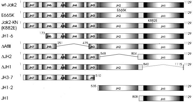
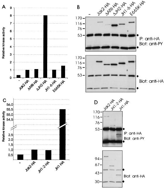
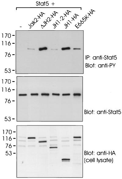
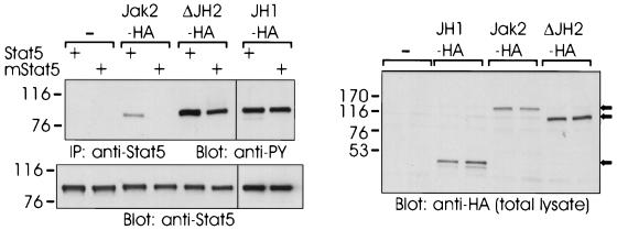
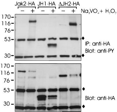
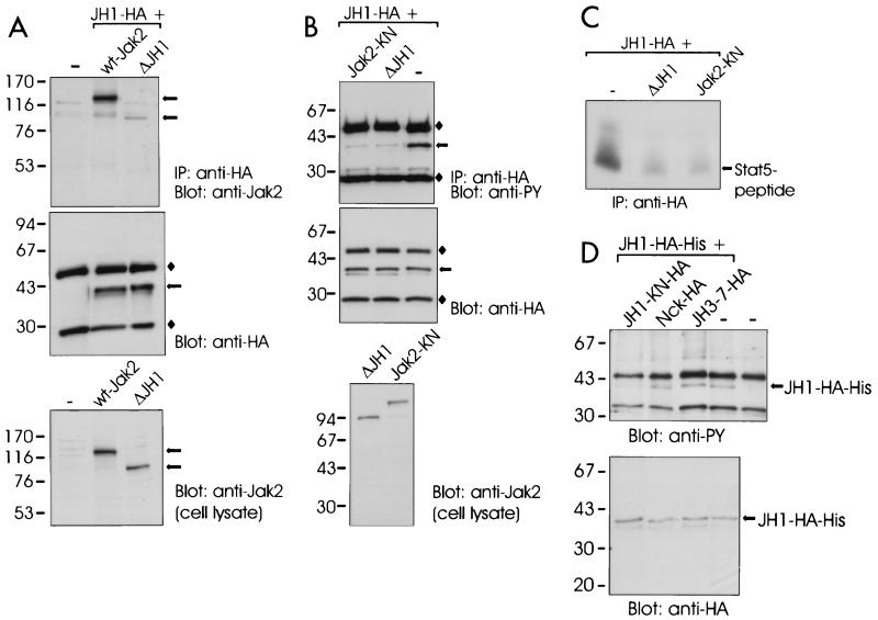
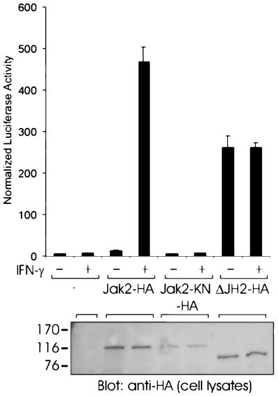
Activation of Jak tyrosine kinases through hematopoietic cytokine receptors occurs as a consequence of ligand-induced aggregation of receptor-associated Jaks and their subsequent autophosphorylation. Jak kinases consist of a C-terminal tyrosine kinase domain, a pseudokinase domain of unknown function, and Jak homology (JH) domains 3 to 7, implicated in receptor-Jak interaction. We analyzed the functional roles of the different protein domains in activation of Jak2. Deletion analysis of Jak2 showed that the pseudokinase domain but not JH domains 3 to 7 negatively regulated the catalytic activity of Jak2 as well as Jak2-mediated activation of Stat5. Phosphorylation of Stat5 by wild-type Jak2 was dependent on the SH2 domain of Stat5; however, this requirement was lost upon deletion of the pseudokinase domain of Jak2. Investigation of the mechanisms of the pseudokinase domain-mediated inhibition of Jak2 suggested that this regulation did not involve protein tyrosine phosphatases. Instead, analysis of interactions between the tyrosine kinase domain and Jak2 suggested that the pseudokinase domain interacted with the kinase domain. Furthermore, coexpression of the pseudokinase domain inhibited the activity of the single tyrosine kinase domain. Finally, deletion of the pseudokinase domain of Jak2 deregulated signal transduction through the gamma interferon receptor by significantly increasing ligand-independent activation of Stat transcription factors. These results indicate that the pseudokinase domain negatively regulates the activity of Jak2, probably through an interaction with the kinase domain, and this regulation is required to keep Jak2 inactive in the absence of ligand stimulation. Furthermore, the pseudokinase domain may have a role in regulation of Jak2-substrate interactions.


Figures

FIG. 1
FIG. 1
Protein products encoded by Jak2 cDNA constructs. Schematic presentation of the proteins encoded by Jak2 deletion constructs. Amino acids encoded by the constructs are indicated; numbers refer to the murine Jak2 sequence. An HA tag, when present, is located in the C terminus and indicated by the suffix HA in the text. The full-length HA-tagged Jak2 is termed Jak2-HA, and the untagged form of Jak2 is termed wt-Jak2.
FIG. 2
FIG. 2
Deletion of JH2 increases the catalytic activity of Jak2. (A) 293T cells were transfected with expression plasmids for Jak2-HA, ΔAflII-HA, ΔJH2-HA, JH1-6-HA, and E665K-HA or left untransfected (—). The lysates were immunoprecipitated with anti-HA antibody, and aliquots were subjected to in vitro kinase assays with Stat1-derived peptide and [γ-32P]ATP as substrates. The peptides were separated by SDS–20% PAGE followed by quantification with a PhosphorImager. Relative catalytic activities are shown. Radioactivity incorporated in the Stat1 peptide by the different Jak2 proteins was normalized to the radioactivity incorporated in the Stat1 peptide by Jak2-HA, which was set at 1. (B) Aliquots of the same immunoprecipitates (IP) were analyzed by SDS–4 to 15% PAGE and blotted with antiphosphotyrosine (anti-PY) or anti-HA antibodies. (C) 293T cells were transfected with expression plasmids for Jak2-HA, JH1-2-HA, and JH1-HA or left untransfected. The lysates were immunoprecipitated with anti-HA antibody and analyzed in in vitro kinase assays as in panel A. (D) Aliquots of the same immunoprecipitates were analyzed by SDS–4 to 15% PAGE and blotted with antiphosphotyrosine or anti-HA antibodies. (B and D) ⧫, immunoglobulin chains. The mobilities of the molecular mass markers (in kilodaltons) are shown on the left.
FIG. 3
FIG. 3
Deletion of JH2 enhances Jak2-mediated activation of Stat5. 293T cells were cotransfected with expression plasmids for Stat5 alone (—) or Stat5 plus Jak2-HA, ΔJH2-HA, JH1-2-HA, JH1-HA, or E665K-HA. Stat5 was immunoprecipitated with anti-Stat5 antibody, and the immunoprecipitates (IP) were analyzed by SDS–7.5% PAGE followed by antiphosphotyrosine (anti-PY) or anti-Stat5 immunoblotting. Aliquots of cell lysates were analyzed by SDS–4 to 15% PAGE followed by anti-HA immunoblotting (lowest panel). The mobilities of the molecular mass markers (in kilodaltons) are shown on the left.
FIG. 4
FIG. 4
SH2 domain of Stat5 required for activation by Jak2 but not by ΔJH2 or JH1. 293T cells were transfected with expression plasmids for Stat5 or SH2 mutant of Stat5 (mStat5). In addition, the cells were transfected with expression plasmids for Jak2-HA, ΔJH2-HA, or JH1-HA or left untransfected (—). Stat5 was immunoprecipitated (IP) with anti-Stat5 antibody. The immunoprecipitates were separated by SDS–7.5% PAGE and blotted with antiphosphotyrosine (anti-PY) or anti-Stat5 antibodies. Aliquots of cell lysates were analyzed by SDS–4 to 15% PAGE followed by anti-HA immunoblotting. The mobilities of the molecular mass markers (in kilodaltons) are shown on the left. Arrows on the right indicate the mobilities of the Jak2 proteins.
FIG. 5
FIG. 5
Effect of PTPase inhibitors on tyrosine phosphorylation of Jak2. 293T cells were transfected with expression plasmids for Jak2-HA, JH1-HA, and ΔJH2-HA. The cells were treated for 30 min with the cell membrane-permeating PTPase inhibitor pervanadate (100 μM) (lanes +) or left untreated (lanes −). The Jak2 proteins were immunoprecipitated (IP) with anti-HA antibody, separated by SDS–4 to 15% PAGE, and immunoblotted with antiphosphotyrosine (anti-PY) or anti-HA antibodies. ⧫, Ig chains. The mobilities of the molecular mass markers (in kilodaltons) are shown on the left.
FIG. 6
FIG. 6
Effect of JH domains on activity of the Jak2 tyrosine kinase domain. (A) 293T cells were transfected with expression plasmids for JH1-HA and untagged wt-Jak2 or ΔJH1 or left untransfected (—). The cells were lysed in Brij 58 buffer, and the lysates were immunoprecipitated (IP) with anti-HA antibody. The immunoprecipitates were separated by SDS–4 to 15% PAGE and immunoblotted with anti-Jak2 or anti-HA antibodies. Aliquots of the cell lysates were analyzed by SDS–7.5% PAGE followed by anti-Jak2 immunoblotting. (B) 293T cells were transfected with JH1-HA expression plasmid alone (lane —) or together with expression plasmids for untagged ΔJH1 or Jak2-KN. JH1-HA was immunoprecipitated (IP) with anti-HA antibody, analyzed by SDS–4 to 15% PAGE, and blotted with antiphosphotyrosine (anti-PY) or anti-HA antibodies. Aliquots of the cell lysates were analyzed by SDS–4 to 15% PAGE followed by anti-Jak2 immunoblotting. ⧫, Ig chains. (C) Aliquots of immunoprecipitates from panel 6B were subjected to in vitro kinase assays with Stat5-derived peptide and [γ-32P]ATP as substrates. The peptides were separated by SDS–20% PAGE followed by autoradiography. (D) 293T cells were transfected with an expression vector for HA- and histidine-tagged JH1 (JH1-HA-His) alone or together with expression plasmids for JH3-7-HA, JH1-KN-HA (K882E in JH1), or Nck-HA. JH1-HA-His was isolated by metal affinity, analyzed by SDS–4 to 15% PAGE, and blotted with antiphosphotyrosine (anti-PY) or anti-HA antibodies. The mobilities of the molecular mass markers (in kilodaltons) are shown on the left in all panels. The arrows on the right indicate the mobilities of the Jak2 proteins.
FIG. 7
FIG. 7
Effect of JH2 on cytokine signal transduction. γ2A cells were transfected with the Stat1-dependent luciferase reporter vector, the pRLTK control vector, and Jak2-HA, Jak2-KN-HA, ΔJH2-HA, or an empty vector (—) as a control. At 5 h after transfection, the cells were removed to serum-free medium and starved for 15 h. The cells were stimulated with IFN-γ (1,000 U/ml) for 5 h or left unstimulated. Luciferase activity was measured as described in Materials and Methods. Shown are the means from three independent experiments and the standard errors of the mean. The mobilities of the molecular mass markers (in kilodaltons) are shown on the left.

References

    1. Andreotti A H, Bunnell S C, Feng S, Berg L J, Schreiber S L. Regulatory intramolecular association in a tyrosine kinase of the Tec family. Nature. 1997;385:93–97. - PubMed
    1. Barahmand-Pour F, Meinke A, Groner B, Decker T. Jak2-Stat5 interactions analyzed in yeast. J Biol Chem. 1998;273:12567–12575. - PubMed
    1. Briscoe J, Rogers N C, Witthuhn B A, Harpur A G, Wilks A F, Stark G R, Ihle J N, Kerr I M. Kinase-negative mutants of JAK1 can sustain interferon-gamma-inducible gene expression but not an antiviral state. EMBO J. 1996;15:799–809. - PMC - PubMed
    1. Cacalano N A, Migone T, Bazan F, Hanson E P, Chen M, Candotti F, O'Shea J J, Johnston J A. Autosomal SCID caused by a point mutation in the N-terminus of Jak3: mapping of the Jak3-receptor interaction domain. EMBO J. 1999;18:1549–1558. - PMC - PubMed
    1. Candotti F, Oakes S A, Johnston J A, Giliani S, Schumacher R F, Mella P, Fiorini M, Ugazio A G, Badolato R, Notarangelo L D, Bozzi F, Macchi P, Strina D, Vezzoni P, Blaese R M, O'Shea J J, Villa A. Structural and functional basis for JAK3-deficient severe combined immunodeficiency. Blood. 1997;90:3996–4003. - PubMed

Publication types

LinkOut - more resources