. 2000 May;20(10):3434-41.
doi: 10.1128/MCB.20.10.3434-3441.2000.

SINE retroposons can be used in vivo as nucleation centers for de novo methylation

Affiliations

SINE retroposons can be used in vivo as nucleation centers for de novo methylation

P Arnaud et al. Mol Cell Biol. 2000 May.

Abstract

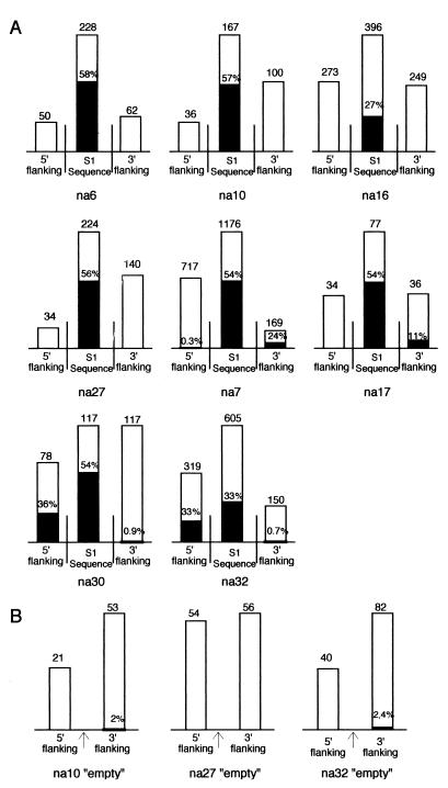
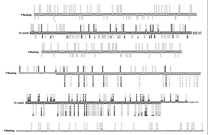
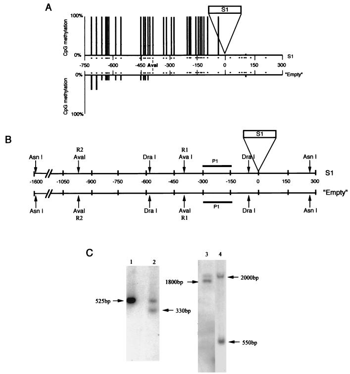
SINEs (short interspersed elements) are an abundant class of transposable elements found in a wide variety of eukaryotes. Using the genomic sequencing technique, we observed that plant S1 SINE retroposons mainly integrate in hypomethylated DNA regions and are targeted by methylases. Methylation can then spread from the SINE into flanking genomic sequences, creating distal epigenetic modifications. This methylation spreading is vectorially directed upstream or downstream of the S1 element, suggesting that it could be facilitated when a potentially good methylatable sequence is single stranded during DNA replication, particularly when located on the lagging strand. Replication of a short methylated DNA region could thus lead to the de novo methylation of upstream or downstream adjacent sequences.


Figures

FIG. 1
FIG. 1
Summary of the methylation status obtained by genomic sequencing of eight S1-containing loci (A) and three corresponding empty loci obtained from an heterozygous plant (B). For each locus (name of the locus indicated below), the height of bars is proportional to the number of cytosines analyzed (indicated above). The proportion of methylated cytosines found in S1 sequences or in upstream or downstream flanking sequences is symbolized by dark portions, and the corresponding percentages (when not 0) are indicated. The arrows for the empty loci indicate the position of insertion of the S1 element in the corresponding S1-containing site.
FIG. 2
FIG. 2
Two detailed examples of genomic sequencing results. (A) Plus and minus strands of the na16 locus. (B) Plus and minus strands of the na32 locus (only the plus strand for the downstream region). Cytosine methylation is indicated by solid circles, while nonmethylated sites are represented by open circles. The S1 sequence is printed in boldface.
FIG. 3
FIG. 3
Evaluation of the upstream methylation spreading for the na32 locus. (A) The methylation status of the empty and S1 allele upstream regions was analyzed by genomic sequencing (upper strand). Only the CpG sites are indicated (by stars). The position of the R1-AvaI site is indicated. The proportion of CpG methylation is represented by bars of different lengths. Nine and seven clones were sequenced, respectively, for the empty and S1 alleles. (B) Restriction map of both alleles. Positions of the probe (P1) and of the two AvaI restriction sites (R1 and R2) are indicated. DraI and AsnI are not sensible for DNA methylation, while AvaI is inhibited by methylation. (C) Southern analysis of the methylation status of the na32 locus. DNA from heterozygous plants were digested with DraI (lane 1), DraI-AvaI (lane 2), AsnI (lane 3), or AsnI-AvaI (lane 4) and probed with the P1 fragment. The sizes of the hybridized fragments (in base pairs) are shown. The size difference between the two fragments revealed after the AsnI digestion (lane 3) can be explained by the presence or absence of the S1 element and confirms the heterozygous state of the plant studied. The R1 site is methylated for the S1 allele but unmethylated for the empty allele. The R2 sites appear to be methylated for the S1 allele.
FIG. 4
FIG. 4
Possible involvement of DNA replication in de novo methylation spreading. Following integration, S1 elements would be targeted for de novo methylation by an unknown mechanism (see text). After replicating the methylated SINE (black boxes), two hemimethylated regions will be formed (black and white boxes), one on the leading strand and one on the lagging strand. DNA methyltransferase (hatched oval) associated with the replication apparatus (grey circle) can recognize these hemimethylated regions and can methylate the corresponding neosynthesized strands (23) (the organization of the replication fork is presented as by Kornberg and Baker [20]). In the 3′ part of the SINE on the lagging strand, the methyltransferase is adjacent to single-stranded DNA. Methylation spreading from the SINE to flanking sequences would depend on the presence on the lagging strand of a potentially good methylatable region, and the polarity of the methylation spreading would depend on the position of replication origin and the direction of the replication fork (A or B). The initial methylation spreading (thicker line) can only concern the immediate S1 flanking region, but since de novo methylation is possible at each round of DNA replication, methylation can potentially spread by this mechanism to sequences thousand of base pairs from the SINEs. Also, we should expect that the methylated version of the locus will rapidly take over the unmethylated one following successive rounds of DNA replication.

References

    1. Bennetzen J L. The mutator transposable element system of maize. Curr Top Microbiol Immunol. 1996;24:195–229. - PubMed
    1. Bird A. The essential of DNA methylation. Cell. 1992;70:5–8. - PubMed
    1. Brutnell T P, Dellaporta S L. Somatic inactivation and reactivation of AC associated with changes in cytosine methylation and transposase expression. Genetics. 1994;138:213–225. - PMC - PubMed
    1. Carotti D, Funiciello S, Palitti F, Strom R. Influence of pre-existing methylation on the de novo activity of eukaryotic DNA methyltransferase. Biochemistry. 1998;37:1101–1108. - PubMed
    1. Chesnokov I, Schmid C W. Flanking sequences of an Alu source stimulate transcription in vitro by interacting with sequence-specific transcription factors. J Mol Evol. 1996;42:30–36. - PubMed

Publication types

LinkOut - more resources