. 2000 May;20(10):3616-25.
doi: 10.1128/MCB.20.10.3616-3625.2000.

UV-Induced stabilization of c-fos and other short-lived mRNAs

Affiliations

UV-Induced stabilization of c-fos and other short-lived mRNAs

C Blattner et al. Mol Cell Biol. 2000 May.

Abstract

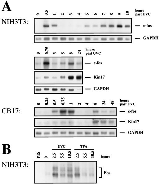
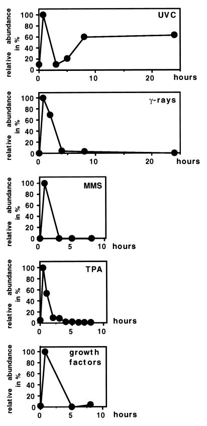
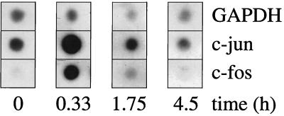
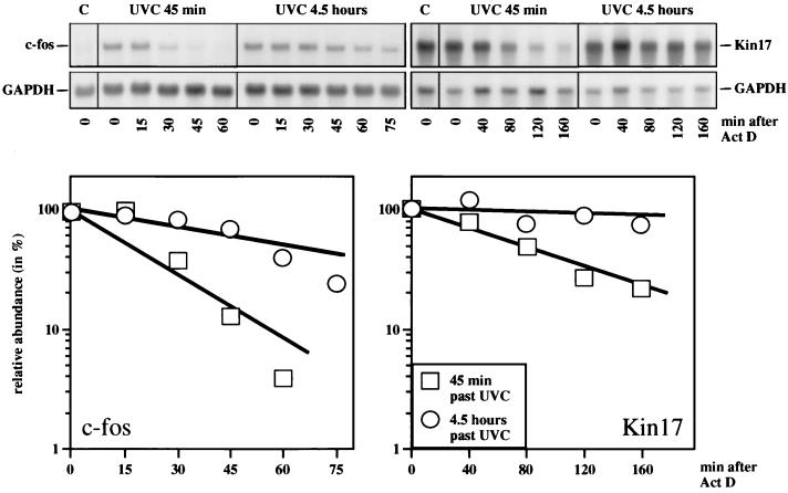
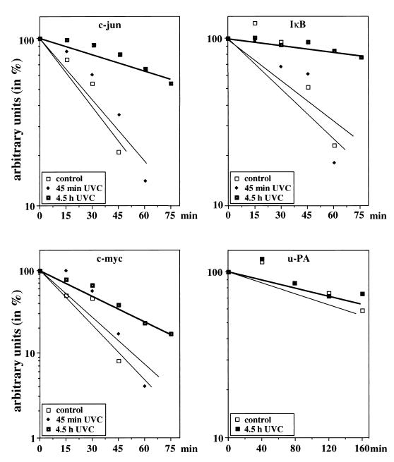
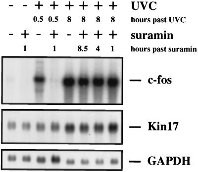
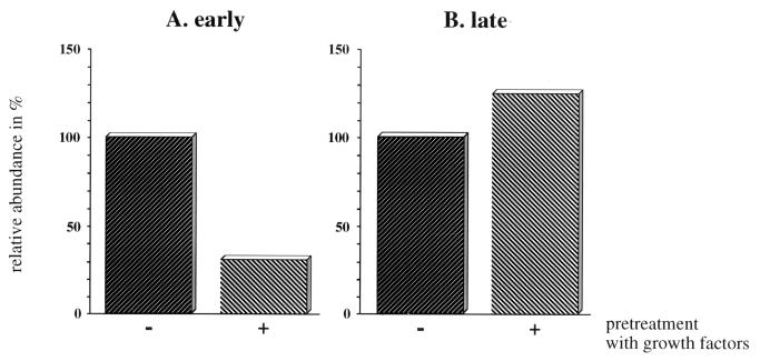
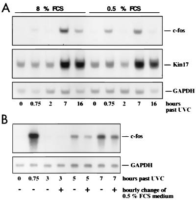
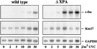
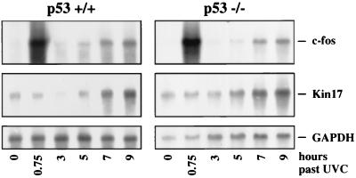
Irradiation of cells with short-wavelength ultraviolet light (UVC) changes the program of gene expression, in part within less than 15 min. As one of the immediate-early genes in response to UV, expression of the oncogene c-fos is upregulated. This immediate induction is regulated at the transcriptional level and is transient in character, due to the autocatalyzed shutoff of transcription and the rapid turnover of c-fos mRNA. In an experiment analyzing the kinetics of c-fos mRNA expression in murine fibroblasts irradiated with UVC, we found that, in addition to the initial transient induction, c-fos mRNA accumulated in a second wave starting at 4 to 5 h after irradiation, reaching a maximum at 8 h, and persisting for several more hours. It was accompanied by an increase in Fos protein synthesis. The second peak of c-fos RNA was caused by an UV dose-dependent increase in mRNA half-life from about 10 to 60 min. With similar kinetics, the mRNAs of other UV target genes (i.e., the Kin17 gene, c-jun, IkappaB, and c-myc) were stabilized (e.g., Kin17 RNA from 80 min to more than 8 h). The delayed response was not due to autocrine cytokine secretion with subsequent autostimulation of the secreting cells or to UV-induced growth factor receptor activation. Cells unable to repair UVC-induced DNA damage responded to lower doses of UVC with an even greater accumulation of c-fos and Kin17 mRNAs than repair-proficient wild-type cells, suggesting that a process in which a repair protein is involved regulates mRNA stability. Although resembling the induction of p53, a DNA damage-dependent increase in p53 was not a necessary intermediate in the stabilization reaction, since cells derived from p53 knockout mice showed the same pattern of c-fos and Kin17 mRNA accumulation as wild-type cells. The data indicate that the signal flow induced by UV radiation addresses not only protein stability (p53) and transcription but also RNA stability, a hitherto-unrecognized level of UV-induced regulation.


Figures

FIG. 1
FIG. 1
Time course of c-fos and Kin17 mRNA induction by UV. (A) NIH 3T3 and CB17 cells were irradiated with UVC (30 J/m2) and harvested at the indicated time points. Poly(A)+ RNA was prepared, and 7.5 μg of each sample was resolved on a 1.4% agarose-formaldehyde gel. The RNA was transferred to a Hybond N+ nylon membrane and sequentially probed with 32P-labelled cDNAs coding for v-fos (hybridizing to c-fos mRNA), Kin17, and GAPDH and placed on X-ray films. (B) NIH 3T3 cells were either irradiated with UVC (30 J/m2) or treated with 60 ng of TPA per ml and harvested 2.5 to 10.5 h after irradiation as indicated. Before harvest, cells were pulse-labelled with 150 μCi of Pro-mix (Amersham) per ml for 2 h and lysed in RIPA buffer. The lysates were precleared by incubation with preimmune serum (PIS) and protein A-agarose. Fos was immunoprecipitated with a polyclonal anti-Fos antibody and protein A-agarose and loaded onto a SDS–10% polyacrylamide gel. The gel was fixed in acetic acid-methanol, enhanced with sodium salicylate, and dried, and X-ray film was exposed for 2 weeks. The bands enclosed within the bracket represent differentially phosphorylated forms of Fos protein.
FIG. 2
FIG. 2
The biphasic induction of c-fos mRNA is specific for UVC irradiation. NIH 3T3 cells were irradiated with UVC (30 J/m2) or gamma rays (50 Gy) or treated with 4 μM MMS, TPA, (60 ng/ml), or a mixture of growth factors (EGF, [2 ng/ml], bFGF [10 ng/ml], and IL-1α [2 ng/ml]). At the indicated time points, poly(A)+ RNA was prepared, and 5 μg of each sample was resolved on a 1.4% agarose-formaldehyde gel. Hybridizations were carried out as described in the legend to Fig. 1. Relative levels normalized for GAPDH were determined by phosphorimager and densitometry.
FIG. 3
FIG. 3
Run-on analysis of c-fos transcription after UV irradiation. NIH 3T3 cells were irradiated with UVC (30 J/m2). At the indicated time points, nuclei were isolated and elongation of transcripts was performed in vitro in the presence of 32P-labelled nucleotide triphosphates. A total of 8 × 10⁵ cpm at each time point were hybridized to plasmids encoding c-fos, c-jun, or GAPDH.
FIG. 4
FIG. 4
UV induces c-fos and Kin17 mRNA stabilization. NIH 3T3 cells were irradiated with UVC (30 J/m2). At 45 min or 4.5 h after irradiation, Act D (5 μg/ml) was added, and the cells were harvested immediately (0) and every 15 min (for determining the decay of c-fos mRNA) or every 40 min (for determining the decay of Kin17 mRNA), respectively (top). Poly(A)+ RNA was prepared, and 15 μg of each sample was resolved and hybridized as described in the legends to Fig. 1 and 2. The specific hybridization signals for c-fos and Kin17 mRNA were corrected for GAPDH mRNA, which was used as a loading control, and plotted in the graphs (bottom). The relative abundance of c-fos and Kin17 mRNA, respectively, at the time of Act D addition was set at 100%.
FIG. 5
FIG. 5
Stabilization of several short-lived mRNAs after UV irradiation. NIH 3T3 cells were irradiated with UVC (30 J/m2) or left unirradiated for control and treated as described in the legend to Fig. 4. Hybond N+ nylon membrane was sequentially probed with 32P-labelled cDNAs encoding c-jun, IκB, c-myc, u-PA, and GAPDH, respectively. Specific hybridization signals were quantified by evaluating scanned X-ray film using NIH Image software, corrected for GAPDH, and plotted. The relative abundance of the particular mRNAs at the time of Act D addition was set at 100%.
FIG. 6
FIG. 6
Pretreatment of cells with suramin does not interfere with c-fos or Kin17 mRNA stabilization. NIH 3T3 cells were grown in DMEM supplemented with 0.5% FCS for 24 h prior to UVC irradiation. The cells were irradiated with UVC (30 J/m2) where indicated or left unirradiated for control. Where marked in the figure, suramin was added to a final concentration of 0.3 mM 30 min prior to UVC irradiation or 4 h (4 h after suramin) or 7 h (1 h past suramin) after irradiation. The cells were harvested at 0.5 h or 8 h after UVC irradiation as shown, poly(A)+ RNA was prepared, and 5 μg of each sample was resolved and hybridized as described in the legends to Fig. 1 and 2.
FIG. 7
FIG. 7
Differential effect of growth factor pretreatment on early and late c-fos induction. NIH 3T3 cells were grown in DMEM supplemented with 0.5% FCS for 24 h prior to UVC irradiation and irradiated with 30 J/m2 UVC. Where marked in the figure, cells were treated with a cocktail of growth factors (EGF, 2 ng/ml; bFGF, 10 ng/ml; IL-1α, 2 ng/ml) at 30 min prior to irradiation. At 45 min (early) (A) and 8 h (late) (B), irradiated cells were harvested. Poly(A)+ RNA was prepared, and 5 μg of each sample was analyzed as described in the legend to Fig. 1. The relative abundance of c-fos mRNA was determined densitometrically and plotted after correction for GAPDH mRNA. The relative abundance of c-fos mRNA in cells that had been irradiated but not treated with growth factor was set at 100%.
FIG. 8
FIG. 8
Influence of serum on RNA stabilization by UV. (A) NIH 3T3 cells were either grown in DMEM supplemented with 8% FCS or were serum starved in DMEM plus 0.5% FCS prior to UVC irradiation for 24 h. Poly(A)+ RNA was analyzed as described in the legends to Fig. 1 and 2. (B) NIH 3T3 cells were serum starved in DMEM supplemented with 0.5% FCS for 24 h prior to UVC irradiation. Cells were irradiated with 30 J/m2 UVC and, where marked in the figure key (+), the culture medium was replaced every hour by fresh DMEM plus 0.5% FCS. Poly(A)+ RNA was prepared and analyzed as described in the legends to Fig. 1 and 2.
FIG. 9
FIG. 9
UV dose dependence of RNA stabilization in cells from XPA knockout mice and from wild-type cells. Fibroblast cell lines from XPA knockout mice (ΔXPA) and from the respective parental mice (wild type) were irradiated with 2, 5, 10, 20, or 30 J/m2 UVC or left unirradiated for control. At 16 h postirradiation, poly(A)+ RNA was prepared and processed as described in the legends to Fig. 1 through 3.
FIG. 10
FIG. 10
Equal induction of RNA stability by UV both in p53+/+ and p53−/− cells. Fibroblast cell lines from p53 knockout mice (p53−/−) and from the wild-type mice (p53+/+) were irradiated with UVC (30 J/m2). The cells were harvested at the indicated time points, and poly(A)+ RNA was prepared and analyzed as described in the legends to Fig. 1 through 3.
FIG. 11
FIG. 11
c-fos is required for cell survival. c-fos+/+, c-fos−/−, and NIH 3T3 cells were irradiated with the indicated UVC dose, counted, and plated in quadruplicate at a density of 103 cells/well in 96-well plates. After 36 h or 5 days, respectively, MTT was added at a final concentration of 1 mg/ml, and the cells were returned to the incubator for additional 4 h. The medium was replaced by isopropanol, and MTT conversion was immediately measured at 590 nm. The mean values of three independent experiments were calculated and plotted as percentages of nonirradiated cells.

References

    1. Adler V, Fuchs S Y, Kim J, Kraft A, King M P, Pelling J, Ronai Z. Jun-NH2-terminal kinase activation mediated by UV-induced DNA lesions in melanoma and fibroblast cells. Cell Growth Differ. 1995;6:1437–1446. - PubMed
    1. Angulo J F, Moreau P L, Maunoury R, Laporte J, Hill A M, Bertolotti R, Devoret R. KIN, a mammalian nuclear protein immunologically related to E. coli RecA protein. Mutat Res. 1989;217:123–134. - PubMed
    1. Angulo J F, Rouer E, Mazin A, Mattei M G, Tissier A, Horellou P, Benarous R, Devoret R. Identification and expression of the cDNA of KIN17, a zinc-finger gene located on mouse chromosome 2, encoding a new DNA-binding protein. Nucleic Acids Res. 1991;19:5117–5123. - PMC - PubMed
    1. Aragane Y, Kulms D, Metze D, Wilkes G, Pöppelmann B, Luger T A, Schwarz T. Ultraviolet light induces apoptosis via direct activation of CD95 (Fas/Apo-1) independently of its ligand CD95L. J Cell Biol. 1998;140:171–182. - PMC - PubMed
    1. Bates S, Vousden K H. Mechanisms of p53-mediated apoptosis. Cell Mol Life Sci. 1999;55:28–37. - PMC - PubMed

Publication types

MeSH terms

LinkOut - more resources