. 2000 May;20(10):3626-32.
doi: 10.1128/MCB.20.10.3626-3632.2000.

Identification of a novel E2F3 product suggests a mechanism for determining specificity of repression by Rb proteins

Affiliations

Identification of a novel E2F3 product suggests a mechanism for determining specificity of repression by Rb proteins

G Leone et al. Mol Cell Biol. 2000 May.

Abstract

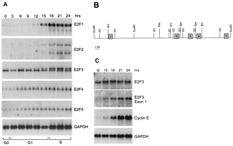
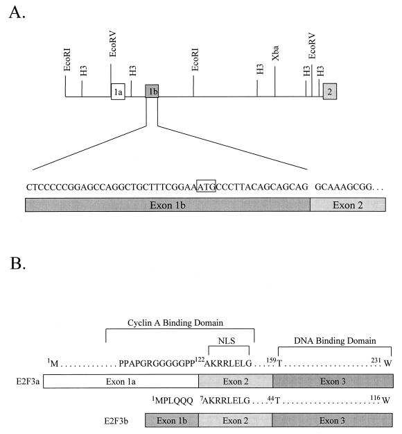
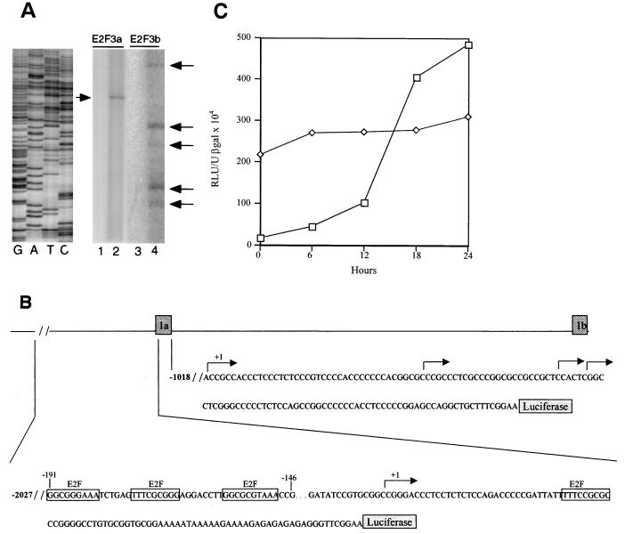
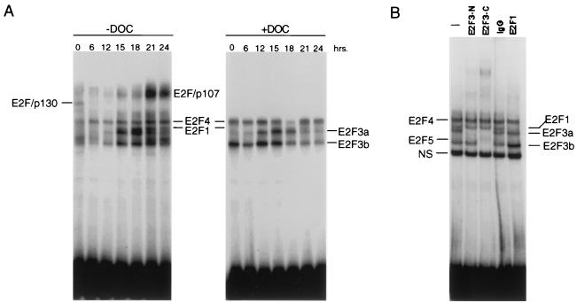
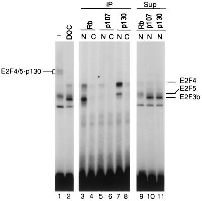
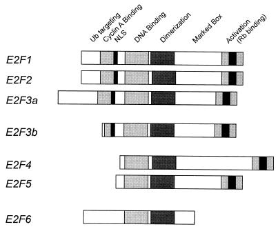
The tumor suppressor function of Rb is intimately related to its ability to interact with E2F and repress the transcription of E2F target genes. Here we describe a novel E2F product that specifically interacts with Rb in quiescent cells. This novel E2F, which we term E2F3b, is encoded by a unique mRNA transcribed from an intronic promoter within the E2F3 locus. The E2F3b RNA differs from the previously characterized E2F3 RNA, which we now term E2F3a, by the utilization of a unique coding exon. In contrast to the E2F3a product that is tightly regulated by cell growth, the E2F3b product is expressed equivalently in quiescent and proliferating cells. But, unlike the E2F4 and E2F5 proteins, which are also expressed in quiescent cells and form complexes with the p130 protein, the E2F3b protein associates with Rb and represents the predominant E2F-Rb complex in quiescent cells. Thus, the previously described specificity of Rb function as a transcriptional repressor in quiescent cells coincides with the association of Rb with this novel E2F product.


Figures

FIG. 1
FIG. 1
Expression of E2F family members following stimulation of cell growth. (A) Expression of E2F family RNAs following stimulation of REF52 cell growth. Cells were brought to quiescence by serum starvation and then stimulated to grow by the addition of fresh media with serum. Samples were taken at the indicated times, and RNA was prepared and analyzed by Northern blotting as described in Materials and Methods. The transition from G1 to S phase, as indicated at the bottom of the figure, was determined by flow cytometry. (B) Genomic organization of the E2F3 locus. The exon-intron structure that includes the initial five exons of the E2F3 locus is shown in relation to restriction maps. (C) Northern analysis with E2F3 exon-specific probes. REF52 cells were brought to quiescence and then stimulated by the addition of serum. Aliquots were taken at the indicated times, and RNA was analyzed as described in Fig. 1. E2F3 RNA was detected with either a full-length cDNA probe (E2F3) or an exon 1-specific probe (E2F3 exon 1a [see Fig. 2]). For comparison, the same blots were probed for expression of the cyclin E gene as well as GAPDH (glyceraldehyde-3-phosphate dehydrogenase) as a loading control.
FIG. 2
FIG. 2
Alternate 5′ exons encode E2F3 RNAs. (A) Genomic structure and organization of the E2F3 locus. The deduced position of the exon 1b, as identified by RACE analysis and sequence comparison, is indicated in relation to the initial E2F3 exon, now termed exon 1a, and in relation to exon 2. The DNA sequence of the longest RACE product is presented. (B) Comparison of the amino acid sequence encoded by E2F3 exon 1a and exon 1b. The sequences homologous to the previously defined cyclin A binding domain of E2F1 are indicated as well as the sequence corresponding to the nuclear localization signal.
FIG. 3
FIG. 3
Cell cycle regulation of the E2F3a and E2F3b promoters. (A) Mapping of E2F3a and E2F3b transcription initiation sites by primer extension analysis. Primers specific for the E2F3a and the E2F3b transcripts (see Materials and Methods) were used with RNA from MEF arrested at G1/S. The major E2F3a extension product (lane 2) and the E2F3b extension products (lane 4) are indicated by arrows. Control reactions lacking MEF RNA are shown in lanes 1 and 3. The size of the primer extension products was compared to a set of DNA sequencing reactions as shown on the left. (B) Genomic organization of the E2F3a and E2F3b promoters. The genomic region including exons 1a and 1b is shown together with DNA sequence immediately upstream of each exon. The positions of transcription start sites, as inferred from the primer extension mapping, are indicated by arrows. In each case, the +1 site represents the longest primer extension product. The site of fusion of each promoter fragment to the luciferase reporter is shown as well as the E2F recognition sites in the E2F3a upstream sequence. (C) Analysis of E2F3a (□) and E2F3b (◊) promoter activity following the stimulation of cell growth. REF52 cells were transfected with 4 μg of E2F3a-luciferase or E2F3b-luciferase, together with 2 μg of the CMV–β-galactosidase vector as an internal control. Transfected cells were brought to quiescence by serum starvation for 48 h and then stimulated by the addition of fresh medium with serum. Samples were taken at the indicated times and assayed for luciferase and β-galactosidase activity. Luciferase activity was normalized to β-galactosidase activity.
FIG. 4
FIG. 4
Control of E2F3 DNA binding activity following stimulation of cell growth. (A) E2F DNA binding activity following stimulation of cell growth. REF52 cells were brought to quiescence and then stimulated by the addition of medium containing 10% serum. Cells were harvested at the indicated times following the stimulation of growth, nuclear extracts were prepared, and E2F DNA binding activity was measured using electrophoretic mobility shift assays as previously described. A sample of each nuclear extract was also treated with DOC and then analyzed for E2F DNA binding activity. (B) Identification of distinct E2F3 activities. A nuclear extract sample from the 18-h time point presented in the left panel (G1/S sample) was incubated without antibody (lane 1), an E2F3 antibody specific for the N terminus of E2F3a (lane 2), and E2F3 C-terminus-specific antibody that recognizes both E2F3a and E2F3b (lane 3), control IgG (lane 4), or an E2F1-specific antibody prior to being subjected to gel shift analysis. The E2F4- and E2F5-specific bands indicated on the DNA gel shift have been identified using specific antibodies against the respective proteins (data not shown).
FIG. 5
FIG. 5
E2F3b is specifically associated with Rb in quiescent cells. Nuclear (N) and cytoplasmic (C) extracts prepared from quiescent cells were immunoprecipitated with antibodies specific for the indicated Rb family member protein. The immunoprecipitates (IP) were treated with DOC to release the associated E2F activities as described in Materials and Methods. The released material was assayed for E2F DNA binding activity by gel mobility shift (lanes 3 to 8). In addition, the supernatants (Sup) from the immunoprecipitations were also assayed for E2F activity (lanes 9 to 11). Finally, a sample of the nuclear extract was directly treated with DOC and then assayed for E2F binding activity (lanes 1 and 2).
FIG. 6
FIG. 6
Relationship of E2F3b to the E2F family. Schematic representation of the entire E2F family with conserved domains indicated.

References

    1. DeGregori J, Kowalik T, Nevins J R. Cellular targets for activation by the E2F1 transcription factor include DNA synthesis and G1/S regulatory genes. Mol Cell Biol. 1995;15:4215–4224. - PMC - PubMed
    1. DeGregori J, Leone G, Miron A, Jakoi L, Nevins J R. Distinct roles for E2F proteins in cell growth control and apoptosis. Proc Natl Acad Sci USA. 1997;94:7245–7250. - PMC - PubMed
    1. Dynlacht B D, Moberg K, Lees J A, Harlow E, Zhu L. Specific regulation of E2F family members by cyclin-dependent kinases. Mol Cell Biol. 1997;17:3867–3875. - PMC - PubMed
    1. Dyson N. The regulation of E2f by pRB-family proteins. Genes Dev. 1998;12:2245–2262. - PubMed
    1. George C P, Kadonaga J T. Primer extension analysis of RNA. In: Krieg P A, editor. A laboratory guide to RNA isolation, analysis, and synthesis. New York, N.Y: Wiley-Liss, Inc.; 1996. pp. 133–139.

Publication types

LinkOut - more resources