. 2000 May;20(10):3667-76.
doi: 10.1128/MCB.20.10.3667-3676.2000.

Distinct phosphoisoforms of the Xenopus Mcm4 protein regulate the function of the Mcm complex

Affiliations

Distinct phosphoisoforms of the Xenopus Mcm4 protein regulate the function of the Mcm complex

I Pereverzeva et al. Mol Cell Biol. 2000 May.

Abstract

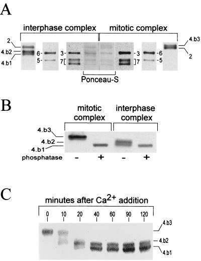
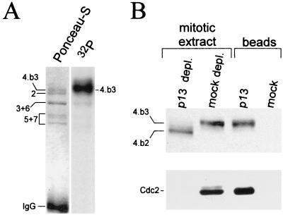
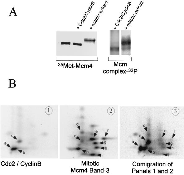
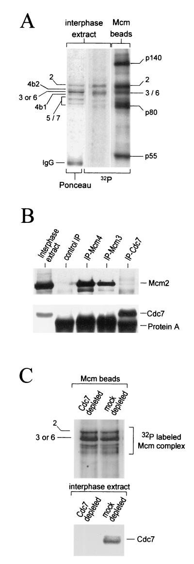
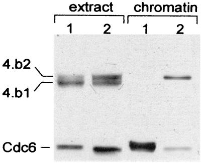
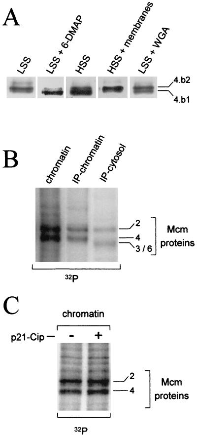
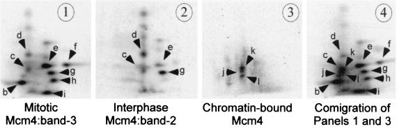
Initiation of DNA replication in eukaryotes requires the assembly of prereplication complexes (pre-Rcs) at the origins of replication. The assembly and function of the pre-Rcs appear to be controlled by phosphorylation events. In this study we report the detailed characterization of the cell cycle phosphorylation of one component of the Xenopus pre-Rcs, the Mcm protein complex. We show that individual Mcm subunits are differentially phosphorylated during the cell cycle. During mitosis, the Mcm4 subunit is hyperphosphorylated, while the other subunits are not actively phosphorylated. The mitotic phosphorylation of Mcm4 requires Cdc2-cyclin B and other unknown kinases. Following exit from mitosis, the Mcm4 subunit of the cytosolic interphase complex undergoes dephosphorylation, and the Mcm2, Mcm3, or Mcm6 subunits are then actively phosphorylated by kinase(s) other than cyclin-dependent kinases (Cdks) or Cdc7. The association of the Mcm complex with the pre-Rcs correlates with the formation of a transient interphase complex. This complex contains an intermediately phosphorylated Mcm4 subunit and is produced by partial dephosphorylation of the mitotic hyperphosphorylated Mcm4 protein. Complete dephosphorylation of the Mcm4 subunit inactivates the Mcm complex and prevents its binding to the chromatin. Once the Mcm complex is assembled on the chromatin the Mcm4 and the Mcm2 proteins are the only subunits phosphorylated during the activation of the pre-Rcs. These chromatin-associated phosphorylations require nuclear transport and are independent of Cdk2-cyclin E. These results suggest that the changes in Mcm4 phosphorylation regulate pre-Rc assembly and the function of the pre-Rcs on the chromatin.


Figures

FIG. 1
FIG. 1
Mitotic and interphase Mcm complexes contain different Mcm4 phosphoisoforms. (A) Western blot analyses of the Mcm subunits composing the interphase and mitotic complexes. Mcm complexes were obtained by immunoprecipitation using anti-Mcm4 antibodies. Each Mcm subunit is referred to by its corresponding number. The three Mcm4 phosphoisoforms are the Mcm4:band-1 (4.b1) and the Mcm4:band-2 (4.b2), present in interphase complexes, and the Mcm4:band-3 (4.b3) present in the mitotic complex. In order to obtain the best resolution between the different Mcm4 isoforms, the Mcm complexes were separated by SDS–7.5% PAGE, run until the 68-kDa molecular mass marker reached the bottom of the gel. (B) Phosphatase treatment of mitotic and interphase extracts. Mcm complexes were incubated with 2 U of alkaline phosphatase for 20 min at 30°C. (C) Kinetics of Mcm4 dephosphorylation following the addition of 0.4 mM CaCl2 to a mitotic extract.
FIG. 2
FIG. 2
Phosphorylation of the Mcm4 subunit of the mitotic Mcm complex is Cdk dependent. (A) The Mcm complex was radiolabeled with 32P in a mitotic extract and then isolated by immunoprecipitation using anti-Mcm4 antibodies. The subunits of the complex were resolved by SDS–7.5% PAGE and transferred to nitrocellulose, and the radiolabeled proteins were visualized with a PhosphorImager. (B) Depletion of Cdks (and their associated proteins) from a mitotic extract was achieved by incubating the extract with an equal volume of p13-Suc1 beads. The beads were recovered by low-speed centrifugation and washed several times with extract buffer containing 0.1% NP-40. The beads and depleted extracts were analyzed for Mcm4 and Cdc2 content by Western blotting using rabbit polyclonal Mcm4 antibodies an a monoclonal anti-Cdc2 antibody (Sc-54; Santa Cruz).
FIG. 3
FIG. 3
Mitotic hyperphosphorylation of the Mcm4 protein requires Cdc2-cyclin B and other kinases. (A) In vitro phosphorylation of Mcm4 by purified Cdc2-cyclin B or a mitotic extract. Phosphorylation reactions were run in kinases buffer containing either 50 U of purified Cdc2-cyclin B or 10 μl of mitotic extract. The substrate was 4 μl of an in vitro-translated [35S]methionine labeled Mcm4 protein or Mcm4 present in an interphase Mcm complex. After a 1-h incubation at room temperature the Mcm proteins were immunoprecipitated with an anti-Mcm4 antibody and separated by SDS-PAGE. The mobility shift of the Mcm4 protein was detected by visualizing both the [35S]methionine label and the incorporated 32P by using a PhosphorImager. The mobility shift of the Mcm4 protein was also confirmed by Western blot analyses (data not shown). (B) Two-dimensional tryptic phosphopeptide mapping was performed on the Mcm4 protein phosphorylated in vitro by Cdc2-cyclin B (panel 1) or in a mitotic extract (panel 2). In panel 3 the two-dimensional phosphopeptide map of a mixture containing samples 1 and 2 is shown. Electrophoretic separation was performed along the horizontal axis, and ascending chromatography was done along the vertical axis.
FIG. 4
FIG. 4
Phosphorylation of the Mcm interphase complex is independent of Cdc7 kinase activity. (A) The Mcm interphase complex was phosphorylated in the extract (lanes 1 and 2) or in vitro by its associated kinase (lane 3). The phosphorylated proteins associated with the immunoprecipitated Mcm complex were separated by SDS-PAGE, transferred to nitrocellulose, and visualized by Ponceau-S staining and autoradiography. (B) Interphase extracts were subjected to immunoprecipitation with antibodies against the Xenopus Mcm3, Mcm4, and Cdc7 proteins, as well as preimmune control antibodies. The immunoprecipitates were resolved by SDS-PAGE and Western blotting using anti-Mcm2 and biotinylated anti-Xenopus Cdc7 antibodies. (C) Interphase extracts were depleted with preimmune (mock) or with anti-Xenopus Cdc7 antibodies bound to protein A-Sepharose beads. Mcm complexes immunoprecipitated from the mock and Cdc7 extracts were then incubated in kinase buffer containing [γ-32P]ATP. After a 1-h incubation at room temperature, the phosphorylated proteins were separated by SDS-PAGE, transferred to nitrocellulose, and visualized by autoradiography.
FIG. 5
FIG. 5
Dephosphorylation of the Mcm4 subunit prevents the binding of the Mcm complex to pre-Rcs. Chromatin binding assays were performed by incubating sperm nuclei (3,000 sperm heads/μl of extract) in interphase Xenopus extract enriched in the dephosphorylated Mcm4:band-1 isoform or in extract containing equivalent amounts of Mcm4:band-2 and Mcm4:band-1. After 15 min of incubation at room temperature, the chromatin was isolated and the presence of Mcm4 and Cdc6 proteins on the chromatin was detected by Western blot analysis.
FIG. 6
FIG. 6
Phosphorylation of the Mcm4 protein on the replicative chromatin. (A) Demembranated sperm nuclei were incubated in a cytosolic high-speed extract (HSS) or interphase extracts (LSS) containing 50 μg of aphidicolin per ml. When indicated, the cytosolic extract was supplemented with a 1/10 volume of a membrane fraction, and the interphase extract was supplemented with either 3 mM DMAP or 0.5 mg of wheat germ agglutinin (WGA) per ml. After a 90-min incubation at room temperature, the chromatin was isolated and the Mcm4 isoform associated with the chromatin was identified by Western blot analysis. (B) Demembranated sperm nuclei were incubated in interphase extracts containing 50 μg of aphidicolin per ml and 1 μCi of [γ-32P]ATP per μl of extract. After a 1-h incubation at room temperature, the chromatin was separated from the cytosol. Mcm proteins were immunoprecipitated from the cytosolic and from the chromatin fractions using anti-Mcm4 antibodies. Western blot analysis and autoradiography identified the phosphorylated Mcm subunits. Total chromatin was also analyzed. (C) Interphase extract was incubated with or without 0.5 μM p21-Cip recombinant protein (40) for 10 min at room temperature. Sperm nuclei, aphidicholin, and [γ-32P]ATP were then added to the extract and incubated for an additional hour. Phosphorylated Mcm proteins on the chromatin were purified as in panel B.
FIG. 7
FIG. 7
Tryptic phosphopeptide maps of the chromatin-bound Mcm4 and cytosolic Mcm4:band-2 isoforms. The phosphorylation of the Mcm protein on the chromatin and of the mitotic Mcm4:band-3 was performed as described in Fig. 6B and 3B, respectively. The 32P-labeled Mcm4:band-2 isoform was obtained by partial dephosphorylation of the mitotic 32P-labeled Mcm4:band-3 following the addition of 0.4 mM CaCl2 to a mitotic extract. Two-dimensional tryptic phosphopeptide mapping of each of the three Mcm4 phosphoisoforms are shown in panel 1 (Mcm4:band-3), panel 2 (Mcm4:band-2), and panel 3 (chromatin-bound Mcm4). Electrophoretic separation was performed along the horizontal axis, and ascending chromatography was done along the vertical axis.

Similar articles

Cited by

References

    1. Aparicio O M, Weinstein D M, Bell S P. Components and dynamics of DNA replication complexes in S. cerevisiae: redistribution of MCM proteins and Cdc45p during S phase. Cell. 1997;91:59–69. - PubMed
    1. Bell S P, Stillman B. ATP-dependent recognition of eukaryotic origins of DNA replication by a multiprotein complex. Nature. 1992;357:128–134. - PubMed
    1. Bousset K, Diffley J F. The Cdc7 protein kinase is required for origin firing during S phase. Genes Dev. 1998;12:480–490. . (Erratum, 12:1072.) - PMC - PubMed
    1. Boyle W J, van der Geer P, Hunter T. Phosphopeptide mapping and phosphoamino acid analysis by two-dimensional separation on thin-layer cellulose plates. Methods Enzymol. 1991;201:110–149. - PubMed
    1. Brown G W, Kelly T J. Purification of Hsk1, a minichromosome maintenance protein kinase from fission yeast. J Biol Chem. 1998;273:22083–22090. - PubMed

Publication types

MeSH terms