. 2000 May;20(10):3685-94.
doi: 10.1128/MCB.20.10.3685-3694.2000.

ADP-ribosylation factor 6 regulates actin cytoskeleton remodeling in coordination with Rac1 and RhoA

Affiliations

ADP-ribosylation factor 6 regulates actin cytoskeleton remodeling in coordination with Rac1 and RhoA

R L Boshans et al. Mol Cell Biol. 2000 May.

Abstract

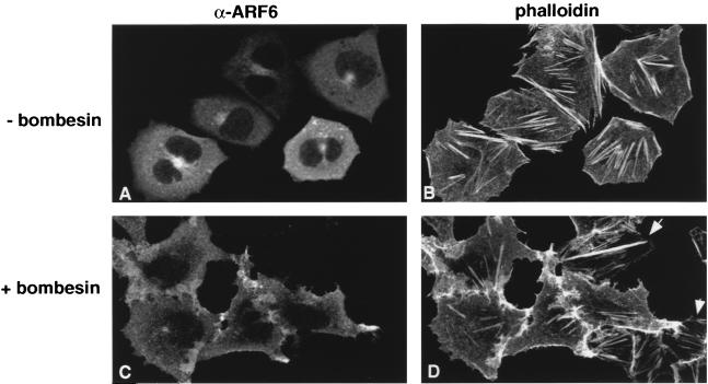
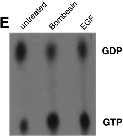
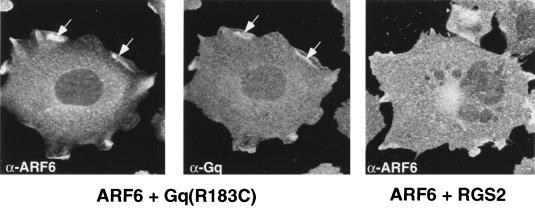
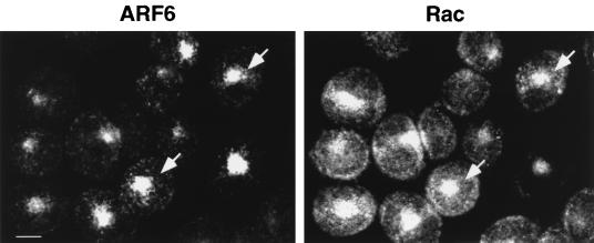
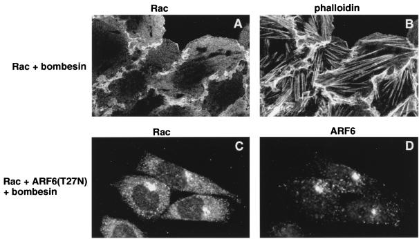
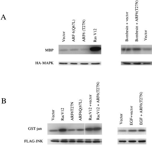
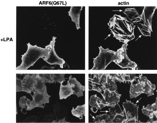
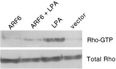
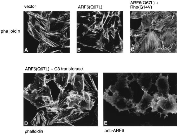
In this study, we have documented an essential role for ADP-ribosylation factor 6 (ARF6) in cell surface remodeling in response to physiological stimulus and in the down regulation of stress fiber formation. We demonstrate that the G-protein-coupled receptor agonist bombesin triggers the redistribution of ARF6- and Rac1-containing endosomal vesicles to the cell surface. This membrane redistribution was accompanied by cortical actin rearrangements and was inhibited by dominant negative ARF6, implying that bombesin is a physiological trigger of ARF6 activation. Furthermore, these studies provide a new model for bombesin-induced Rac1 activation that involves ARF6-regulated endosomal recycling. The bombesin-elicited translocation of vesicular ARF6 was mimicked by activated Galphaq and was partially inhibited by expression of RGS2, which down regulates Gq function. This suggests that Gq functions as an upstream regulator of ARF6 activation. The ARF6-induced peripheral cytoskeletal rearrangements were accompanied by a depletion of stress fibers. Moreover, cells expressing activated ARF6 resisted the formation of stress fibers induced by lysophosphatidic acid. We show that the ARF6-dependent inhibition of stress fiber formation was due to an inhibition of RhoA activation and was overcome by expression of a constitutively active RhoA mutant. The latter observations demonstrate that activation of ARF6 down regulates Rho signaling. Our findings underscore the potential roles of ARF6, Rac1, and RhoA in the coordinated regulation of cytoskeletal remodeling.


Figures

FIG. 1
FIG. 1
Bombesin promotes ARF6 activation. (A to D) Bombesin redistributes ARF6 from perinuclear vesicles to the plasma membrane. Cells transfected with wild-type ARF6 were treated without (A and B) or with (C and D) 15 nM bombesin for 15 min at 37°C. Cells were fixed and labeled with affinity-purified anti-ARF6 polyclonal antibody and were processed for immunofluorescence microscopy. Cytoskeletal rearrangements were visualized by rhodamine phalloidin staining. Untransfected cells in panel D that exhibit stress fibers are indicated by arrows. (E) GTP loading of ARF6 in intact cells. CHO cells treated with or without bombesin or EGF as indicated were transfected with HA-tagged ARF6 and were labeled with [32P]orthophosphate. ARF6 was immunoprecipitated with anti-HA monoclonal antibody, and bound nucleotides were eluted, separated by thin-layer chromatography, and subjected to autoradiography. The data shown are representative of three separate experiments.
FIG. 1
FIG. 1
Bombesin promotes ARF6 activation. (A to D) Bombesin redistributes ARF6 from perinuclear vesicles to the plasma membrane. Cells transfected with wild-type ARF6 were treated without (A and B) or with (C and D) 15 nM bombesin for 15 min at 37°C. Cells were fixed and labeled with affinity-purified anti-ARF6 polyclonal antibody and were processed for immunofluorescence microscopy. Cytoskeletal rearrangements were visualized by rhodamine phalloidin staining. Untransfected cells in panel D that exhibit stress fibers are indicated by arrows. (E) GTP loading of ARF6 in intact cells. CHO cells treated with or without bombesin or EGF as indicated were transfected with HA-tagged ARF6 and were labeled with [32P]orthophosphate. ARF6 was immunoprecipitated with anti-HA monoclonal antibody, and bound nucleotides were eluted, separated by thin-layer chromatography, and subjected to autoradiography. The data shown are representative of three separate experiments.
FIG. 2
FIG. 2
Effect of Gαq(R183C) and RGS2 on ARF6 distribution. Cells cotransfected with plasmids encoding HA-tagged wild-type ARF6 and Gαq(R183C) were fixed and labeled with anti-HA monoclonal antibody (left) and affinity-purified anti-Gαq polyclonal antibody (middle) and were processed for immunofluorescence. Cells cotransfected with plasmids encoding ARF6 and RGS2-GFP were treated with 15 nM bombesin for 15 min and were fixed and labeled with anti-ARF6 antibody (right). RGS2 expression was monitored by GFP (data not shown). Wild-type ARF6 and Gαq colocalize at the cell surface (arrows), whereas coexpression of RGS2 inhibits the redistribution of ARF6 to the plasma membrane.
FIG. 3
FIG. 3
Overlapping subcellular distribution of ARF6 and Rac1 in CHO cells. Cells coexpressing wild-type ARF6 and Rac1 were fixed and processed for immunofluorescence by using affinity-purified antibodies against ARF6 and Rac1. ARF6 localizes predominantly to the perinuclear region of the cell. Rac1 colocalizes with ARF6 in perinuclear vesicles (arrows) but also exhibits a diffuse cytosolic staining and plasma membrane labeling.
FIG. 4
FIG. 4
Bombesin-induced redistribution of Rac1 to the plasma membrane is inhibited by ARF6(T27N). Cells expressing wild-type Rac1 (A and B) or Rac1 plus ARF6(T27N) (C and D) were treated with 15 nM bombesin for 15 min at 37°C and were fixed and processed for indirect immunofluorescence. Cells expressing Rac1 were labeled with anti-Rac1 monoclonal antibody (A) and with rhodamine phalloidin (B). Cells coexpressing Rac1 and ARF6(T27N) were labeled with anti-Rac1 monoclonal antibody (C) and anti-ARF6 rabbit polyclonal antibody (D). As shown, Rac1 localized to the lamellipodia induced on bombesin treatment. Note the formation of stress fibers in the cells. Coexpression of ARF6(T27N) prevents the redistribution of Rac1 to the plasma membrane upon bombesin treatment.
FIG. 5
FIG. 5
(A) Effects of ARF6 on MAPK kinase activity. CHO cells were cotransfected with 5 μg of HA-tagged MAPK and 5 μg of the indicated constructs. Cells in lanes 1 and 2 were treated with bombesin (0.2 μM). HA-tagged MAPK was isolated from cell lysates by immunoprecipitation with anti-HA monoclonal antibody 12CA5, and MAPK activity was measured in an immunocomplex kinase assay with MBP as a substrate. Radioactivity incorporated into MBP was visualized by autoradiography. Expression of MAPK was determined by protein immunoblot analysis by using anti-HA antibodies and was found to be similar in each sample. (B) Effects of ARF6 on JNK activity. CHO cells were cotransfected with 5 μg of HA-tagged-JNK1 and 5 μg of the indicated constructs. Cells in lane 2 and 3 were treated with EGF (50 ng/ml). JNK activity was measured by immunocomplex kinase assays using GST-Jun as substrate and was visualized by autoradiography. Expression of JNK1 was determined by Western blot analysis by using anti-HA antibodies and was found to be similar in each sample.
FIG. 6
FIG. 6
Effect of LPA treatment on stress fiber formation in ARF6(Q67L)-expressing cells. ARF6-expressing serum-starved cells were treated with 1 μM LPA for 10 min at 37°C and were fixed and labeled with anti-ARF6 antibodies and phalloidin. ARF6(Q67L)-expressing cells were resistant to stress fiber formation, whereas untransfected cells (arrow) exhibited stress fibers in response to LPA.
FIG. 7
FIG. 7
Binding of Rho-GTP to RBD-GST. Lysates of cells transfected with expression plasmids encoding indicated proteins were incubated with RBD-GST, and bound Rho-GTP was analyzed by Western blotting with anti-RhoA polyclonal antibodies (upper panel). Cells lysates were resolved in SDS gels and were immunoblotted for total Rho with anti-RhoA antibodies (lower panel).
FIG. 8
FIG. 8
Effect of Rho(G14V) and C3 transferase on ARF6(Q67L)-mediated cytoskeletal rearrangements. Untransfected cells (A), cells transfected with ARF6(Q67L) (B), cells cotransfected with ARF6(Q67L) and Rho(G14V) (C), or cells microinjected with 0.3 mg of myrARF6(Q67L) per ml and 100 μg of C3 transferase per ml (D and E) were fixed and labeled with rhodamine phalloidin to visualize actin filament distribution. Rho(G14V) induces stress fiber formation in ARF6(Q67L)-expressing cells, and C3 transferase did not inhibit ARF6-mediated cytoskeletal rearrangements. Identification of microinjected cells was confirmed by labeling with affinity-purified anti-ARF6 rabbit polyclonal antisera (E).

References

    1. Aprikian A G, Tremblay L, Han K, Chevalier S. Bombesin stimulates the motility of human prostate-carcinoma cells through tyrosine phosphorylation of focal adhesion kinase and of integrin-associated proteins. Int J Cancer. 1997;72:498–504. - PubMed
    1. Bretscher M S. Getting membrane flow and the cytoskeleton to cooperate in moving cells. Cell. 1996;87:601–606. - PubMed
    1. Bretscher M S, Aguado-Velasco C. EGF induces recycling membrane to form ruffles. Curr Biol. 1998;8:721–724. - PubMed
    1. Brown H A, Gutowski S, Moomaw C R, Slaughter C, Sternweiss P C. ADP-ribosylation factor, a small GTP-dependent regulatory protein stimulates phospholipase D activity. Cell. 1993;75:1137–1144. - PubMed
    1. Charlesworth A, Rozengurt E. Bombesin and neuromedin B stimulate the activation of p42(mapk) and p74(raf-1) via a protein kinase C-independent pathway in Rat-1 cells. Oncogene. 1997;14:2323–2329. - PubMed

Publication types

MeSH terms