. 2000 May 9;97(10):5616-20.
doi: 10.1073/pnas.090034797.

Sodium channel Na(v)1.6 is localized at nodes of ranvier, dendrites, and synapses

Affiliations

Sodium channel Na(v)1.6 is localized at nodes of ranvier, dendrites, and synapses

J H Caldwell et al. Proc Natl Acad Sci U S A. .

Abstract

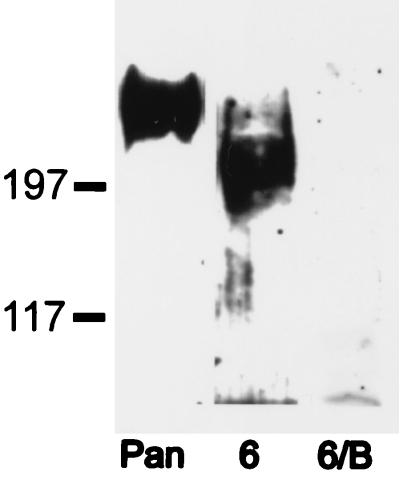
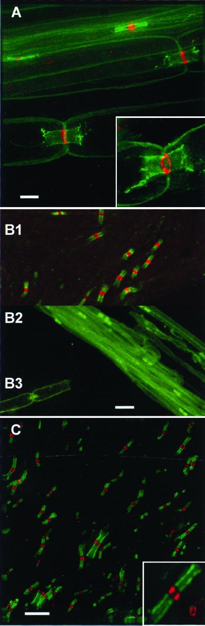
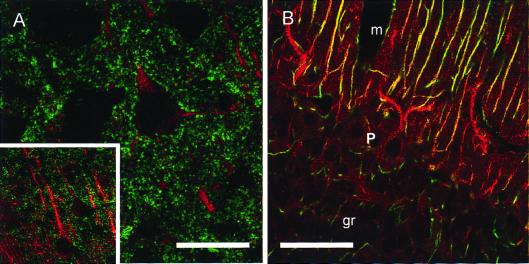
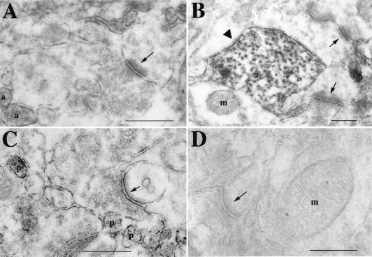
Voltage-gated sodium channels perform critical roles for electrical signaling in the nervous system by generating action potentials in axons and in dendrites. At least 10 genes encode sodium channels in mammals, but specific physiological roles that distinguish each of these isoforms are not known. One possibility is that each isoform is expressed in a restricted set of cell types or is targeted to a specific domain of a neuron or muscle cell. Using affinity-purified isoform-specific antibodies, we find that Na(v)1.6 is highly concentrated at nodes of Ranvier of both sensory and motor axons in the peripheral nervous system and at nodes in the central nervous system. The specificity of this antibody was also demonstrated with the Na(v)1.6-deficient mouse mutant strain med, whose nodes were negative for Na(v)1.6 immunostaining. Both the intensity of labeling and the failure of other isoform-specific antibodies to label nodes suggest that Na(v)1.6 is the predominant channel type in this structure. In the central nervous system, Na(v)1.6 is localized in unmyelinated axons in the retina and cerebellum and is strongly expressed in dendrites of cortical pyramidal cells and cerebellar Purkinje cells. Ultrastructural studies indicate that labeling in dendrites is both intracellular and on dendritic shaft membranes. Remarkably, Na(v)1.6 labeling was observed at both presynaptic and postsynaptic membranes in the cortex and cerebellum. Thus, a single sodium channel isoform is targeted to different neuronal domains and can influence both axonal conduction and synaptic responses.


Figures

Figure 1
Figure 1
Immunoblot of rat brain membranes. A sodium channel pan-specific antibody labeled a major band at ≈250 kDa (pan lane). An anti-Nav1.6 antibody labeled a band at ≈225 kDa (6 lane). Nav1.6 labeling was abolished by preincubation of the antibody with the antigenic peptide (6/B lane).
Figure 2
Figure 2
Node of Ranvier labeling in the PNS (rat and mouse sciatic) and CNS (rat optic nerve). Immunofluorescence with anti-Nav1.6 antibody (red) and anti-caspr (green). Anti-caspr labeled the paranodal region on either side of the nodal labeling by anti-Nav1.6. (A) Rat sciatic nerve. All nodes labeled by caspr were labeled for Nav1.6. (A Inset) Another node viewed obliquely such that the circumferential labeling of axonal membrane can be seen. (B) Mouse sciatic nerve. Labeling controls for Nav1.6 and caspr. (B1) Wild-type mouse labeled for Nav1.6 (red) at nodes shown by caspr labeling (green). (B2) Mutant mouse (medJ) lacking Nav1.6 had no labeling for Nav1.6 at nodes. (B3) One node illustrating that preincubation of the anti-Nav1.6 antibody with the peptide eliminated nodal labeling in the wild-type mouse. (C) Optic nerve axons. Optical sections (0.2 μm apart; n = 25) were deconvolved and projected onto a single plane. Every node was labeled for Nav1.6 (caspr labeling in the figure without Nav1.6 labeling was at the top or bottom of the series of sections). (C Inset) Two nodes, one viewed longitudinally and one viewed transversely such that the nodal labeling appears circular. (Bars = 5 μm for A and C, 3.3 μm for A Inset and C Inset, 6 μm for B1, 10 μm for B2, and 13 μm for B3.)
Figure 3
Figure 3
Immunofluorescence labeling of cerebral cortex for Nav1.6 and synaptic vesicles and of cerebellar cortex for Nav1.6 and glial cells. Single confocal sections. (A) Cerebral cortex labeled for Nav1.6 (red) and SV2 (green). Apical dendrites of neurons were labeled for Nav1.6 within the dendrite. SV2 labeling was throughout the neuropil and outlined the cell bodies and large dendrites of the neurons. (Inset) Deconvolved image of cortical neurons (lower magnification than in A). (B) Cerebellar cortex labeled for Nav1.6 (red) and GFAP (green). Nav1.6 labeling was intense in apical dendrites of Purkinje cells in the vertically oriented GFAP-positive radial glial cell processes (yellow) and was diffusely present in the molecular layer (m). Glial cell processes that were GFAP-positive in the granule cell layer (gr) were not labeled for Nav1.6. Purkinje cell bodies (P layer) were lightly labeled in the cytoplasm. (Bars = 20 μm for A, 75 μm for Inset, and 50 μm for B.)
Figure 4
Figure 4
Axonal and dendritic labeling of Nav1.6 in cerebral and cerebellar cortex. (A) Labeling of presynaptic and postsynaptic membranes (arrow) in cerebral cortex. Plasma membranes of axons (a) were also labeled. (B) Synaptic membrane labeling (arrows) as well as membrane (arrowhead) and microtubule-associated labeling in a dendrite of cerebral cortex. Plasma membrane labeling of the dendrite was independent of a synapse, and adjacent membrane (arrowhead) was unlabeled, indicating that labeling of adjacent membranes at synapses was not due to diffusion of reaction product. A mitochondrion (m) was unlabeled. (C) Molecular layer of the cerebellar cortex. Presynaptic and postsynaptic labeling between a parallel fiber (with synaptic vesicles) and a dendritic spine (arrow) of a Purkinje cell. Parallel fiber axon profiles (p) were also labeled. (D) Cerebral cortex showed no labeling when the antibody was preincubated with the antigenic peptide. A synapse (arrow) and mitochondrion (m) were not labeled. (Bars = 0.4 μm for A, C, and D and 0.3 μm for B.)

Similar articles

Cited by

References

    1. Dugandzija-Novakovic S, Koszowski A G, Levinson S R, Shrager P. J Neurosci. 1995;15:492–503. - PMC - PubMed
    1. Kaplan M R, Meyer-Franke A, Lambert S, Bennett V, Duncan I D, Levinson S R, Barres B A. Nature (London) 1997;386:724–728. - PubMed
    1. Salzer J L. Neuron. 1999;23:627–629. - PubMed
    1. Huguenard J R, Hamill O P, Prince D A. Proc Natl Acad Sci USA. 1989;86:2473–2477. - PMC - PubMed
    1. Regehr W G, Armstrong C M. Curr Biol. 1994;4:436–439. - PubMed

Publication types

MeSH terms