. 2000 May 9;97(10):5428-33.
doi: 10.1073/pnas.090091197.

Preservation of myocardial beta-adrenergic receptor signaling delays the development of heart failure after myocardial infarction

Affiliations

Preservation of myocardial beta-adrenergic receptor signaling delays the development of heart failure after myocardial infarction

D C White et al. Proc Natl Acad Sci U S A. .

Abstract

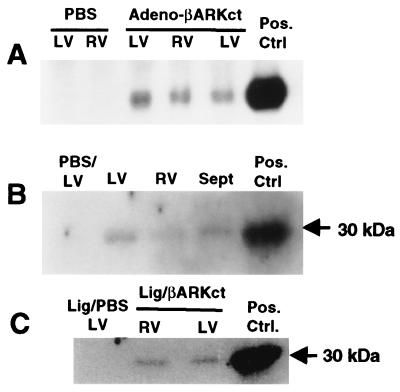
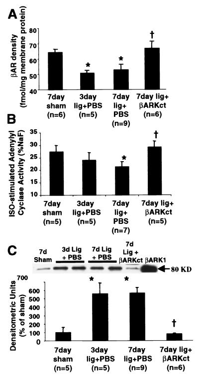
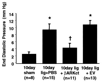
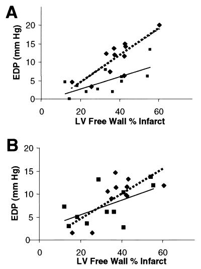
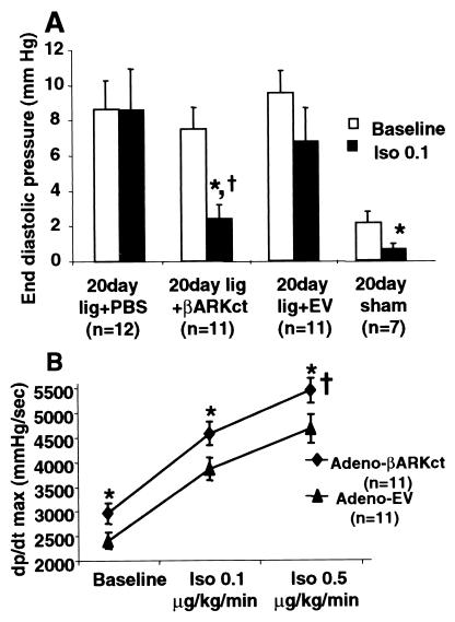
When the heart fails, there is often a constellation of biochemical alterations of the beta-adrenergic receptor (betaAR) signaling system, leading to the loss of cardiac inotropic reserve. betaAR down-regulation and functional uncoupling are mediated through enhanced activity of the betaAR kinase (betaARK1), the expression of which is increased in ischemic and failing myocardium. These changes are widely viewed as representing an adaptive mechanism, which protects the heart against chronic activation. In this study, we demonstrate, using in vivo intracoronary adenoviral-mediated gene delivery of a peptide inhibitor of betaARK1 (betaARKct), that the desensitization and down-regulation of betaARs seen in the failing heart may actually be maladaptive. In a rabbit model of heart failure induced by myocardial infarction, which recapitulates the biochemical betaAR abnormalities seen in human heart failure, delivery of the betaARKct transgene at the time of myocardial infarction prevents the rise in betaARK1 activity and expression and thereby maintains betaAR density and signaling at normal levels. Rather than leading to deleterious effects, cardiac function is improved, and the development of heart failure is delayed. These results appear to challenge the notion that dampening of betaAR signaling in the failing heart is protective, and they may lead to novel therapeutic strategies to treat heart disease via inhibition of betaARK1 and preservation of myocardial betaAR function.


Figures

Figure 1
Figure 1
βARKct expression after in vivo adenoviral-mediated intracoronary transgene delivery of 5 × 10¹¹ tvp Adeno-βARKct. (A) Northern analysis of βARKct mRNA from total RNA isolated from representative Adeno-βARKct and PBS-treated rabbit hearts. (B) βARKct peptide expression in the normal heart as visualized via a Western blot of representative cardiac extracts from Adeno-βARKct and PBS-treated rabbit hearts. (C) Representative Western blot of extracts from infarcted rabbit hearts that received PBS or Adeno-βARKct before LCx ligation. The positive control was RNA or protein isolated from cultured cardiomyocytes infected with Adeno-βARKct.
Figure 2
Figure 2
βAR signaling after LCx ligation and treatment with either PBS or 5 × 10¹¹ tvp Adeno-βARKct. (A) βAR density assessed in purified myocardial membranes and expressed (mean ± SEM) as fmol per milligram of membrane protein. *, P < 0.05 vs. sham; †, P < 0.05 vs. PBS (Student's t test). (B) Myocardial membrane Iso-stimulated adenylyl cyclase activity presented (mean ± SEM) as pmol cAMP/min/mg protein and normalized to the activity (%) stimulated by 10 mM NaF. *, P < 0.05 vs. sham; †, P < 0.05 vs. PBS (Student's t test). (C) Myocardial βARK1 protein levels determined by protein immunoblotting of βARK1 immunoprecipitated cardiac cytosolic extracts. (Upper) Representative Western blot for the indicated treatment groups by using purified βARK1 as a positive control. The histograms show the mean ± SEM. *, P < 0.001 vs. sham; †, P < 0.001 vs. PBS (Student's t test).
Figure 3
Figure 3
LV EDP 10 days after LCx ligation and treatment with either PBS, 5 × 10¹¹ tvp EV, or 5 × 10¹¹ tvp Adeno-βARKct. *, P < 0.001 vs. sham; †, P < 0.05 vs. lig + PBS and vs. lig + EV (one-way ANOVA with Newman–Keuls post-hoc analysis).
Figure 4
Figure 4
Baseline EDP is plotted vs. infarct size for EV (n = 11; diamonds, dotted line) and Adeno-βARKct-treated (n = 11; squares, solid line) rabbits. (A) At day 10, the equations for the regression lines are y = 0.3617x −2.725, r = 0.81 for EV, and y = 0.1663x −0.6297, r = 0.68 for Adeno-βARKct; P < 0.005 for overall coincidence, indicating that the lines are significantly different. (B) At day 20, y = 0.1580x + 2.333, r = 0.57 for EV and y = 0.2719x −0.7664, r = 0.72 for Adeno-βARKct, and the lines are not significantly different (P = 0.50).
Figure 5
Figure 5
Hemodynamic function 20 days after LCx ligation and treatment with either PBS, 5 × 10¹¹ tvp EV or 5 × 10¹¹ tvp Adeno-βARKct. (A) LV EDP values at baseline (open bars) and in response to Iso (closed bars) (Iso, 0.1 μg/kg/min). *, P < 0.01 vs. baseline values (paired t test); †, P < 0.05 vs. Iso–EV (Student's t test). There was no significant response to Iso in the PBS or EV groups. (B) LV dP/dtmax values at baseline and in response to Iso in Adeno-βARKct-treated (diamonds, n = 11) compared with EV-treated (triangles, n = 11) infarcted rabbits. †, P < 0.05 (ANOVA with repeated measures); *, P < 0.01 for individual doses (ANOVA with Newman–Keuls post hoc analysis).

References

    1. Brodde O E. Pharmacol Ther. 1993;60:405–430. - PubMed
    1. Leimbach W N, Wallin G, Victor R G, Aylward P E, Sundlof G, Mark A L. Circulation. 1986;73:913–919. - PubMed
    1. Bristow M R, Ginsburg R, Minobe W, Cubicciotti R, Sageman W S, Lurie K, Billingham M E, Harrison D C, Stinson E B. N Engl J Med. 1982;307:205–211. - PubMed
    1. Bristow M R, Minobe W, Raynolds M V, Port J D, Rasmussen R, Ray P E, Feldman A M. J Clin Invest. 1993;92:2737–2745. - PMC - PubMed
    1. Ungerer M, Bohm M, Elce J S, Erdmann E, Lohse M L. Circulation. 1993;87:454–463. - PubMed

Publication types

MeSH terms

Substances

LinkOut - more resources