. 2000 Apr;129(8):1543-52.
doi: 10.1038/sj.bjp.0703145.

Upregulation by glucocorticoids of responses to eosinopoietic cytokines in bone-marrow from normal and allergic mice

Affiliations

Upregulation by glucocorticoids of responses to eosinopoietic cytokines in bone-marrow from normal and allergic mice

M I Gaspar Elsas et al. Br J Pharmacol. 2000 Apr.

Abstract

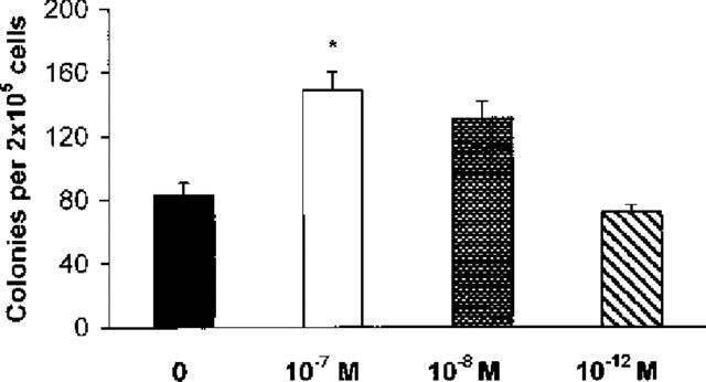
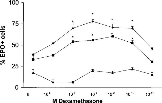
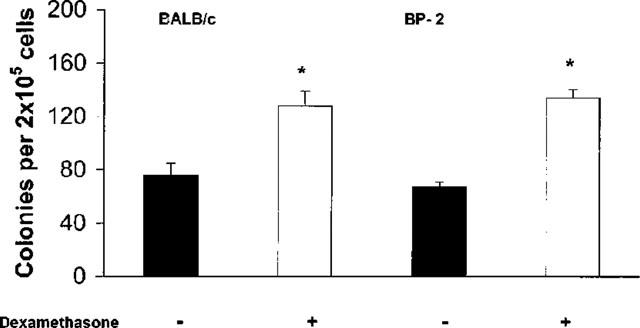
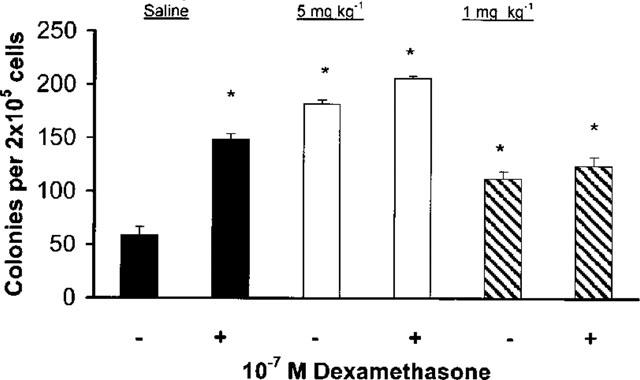
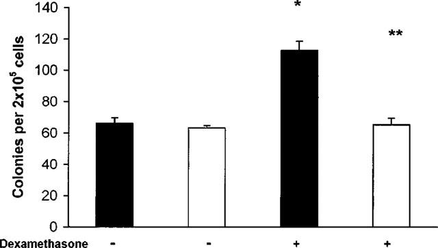
Since the production of eosinopoietic cytokines (GM-CSF, IL-3, IL-5) is inhibited by glucocorticoids, while responsiveness to these cytokines is enhanced in bone-marrow of allergic mice, we studied the ability of glucocorticoids to modulate murine bone-marrow eosinopoiesis. Progenitor (semi-solid) and/or precursor (liquid) cultures were established from bone-marrow of: (a) normal mice; (b) ovalbumin-sensitized and challenged mice or (c) dexamethasone (1-5 mg kg(-1)) injected mice. Cultures were established with GM-CSF (2 ng ml(-1)) or IL-5 (1 ng ml(-1)), respectively, alone or associated with dexamethasone, hydrocortisone or corticosterone. Total myeloid colony numbers, frequency and size of eosinophil colonies, and numbers of eosinophil-peroxidase-positive cells were determined at day 7. In BALB/c mice, dexamethasone (10(-7) M) increased GM-CSF-stimulated myeloid colony formation (P = 0.01), as well as the frequency (P=0.01) and size (P<0.01) of eosinophil colonies. Dexamethasone (10(-7) M) alone had no effect. Dexamethasone (10(-7)-10(-10) M) increased (P<0.002) eosinophil precursor responses to IL-5. Potentiation by dexamethasone was still detectable: (a) on low density, immature, nonadherent BALB/c bone-marrow cells, (b) on bone-marrow from other strains, and (c) on cells from allergic mice. Hydrocortisone and corticosterone had similar effects. Dexamethasone administered in vivo, 24 h before bone-marrow harvest, increased subsequent progenitor responses to GM-CSF (P = 0.001) and precursor responses to IL-5 (P<0.001). These effects were blocked by RU 486 (20 mg kg(-1), orally, 2 h before dexamethasone, or added in vitro at 10 microM, P<0.001). Glucocorticoids, acting in vivo or in vitro, through glucocorticoid receptors, enhance bone-marrow eosinopoiesis in naïve and allergic mice.


Figures

Figure 1
Figure 1
Effect of dexamethasone on GM-CSF-induced colony formation. The data are mean±s.e.mean of the number of myeloid colonies formed by bone-marrow from naïve BALB/c mice. GM-CSF (2 ng ml−1) alone, closed bar. GM-CSF in association with dexamethasone, 10−7M, open bar, 10−8M, stippled bar, 10−12M, cross-hatched bar. Data are derived from a total of 24 experiments, each experiment performed with pooled bone-marrow cells from 5–7 mice. Asterisk indicates significant difference relative to the GM-CSF control (P<0.001).
Figure 2
Figure 2
Effect of in vitro exposure to dexamethasone on IL-5-driven eosinophil differentiation in liquid culture. Data are mean±s.e.mean of the per cent EPO+ cells in liquid cultures established from naïve BALB/c mice, in the presence of 1 (losenges), 0.1 (squares) or 0.01 (triangles) ng ml−1 IL-5 and in the absence (0) or in the presence of dexamethasone, at the indicated molar concentrations. Data are derived from four experiments. Asterisks indicate significant differences relative to the respective IL-5 controls (P<0.002 for dexamethasone at 10−7–10−10M in the upper curve, P<0.05 for the same concentration range in the middle curve).
Figure 3
Figure 3
Morphological features of EPO+ cells grown in the presence of dexamethasone plus IL-5. Cytocentrifuge smears from 7 day-liquid bone-marrow cultures established in the presence of 1 ng ml−1 IL-5 (a and c) or of IL-5 associated with 10−7M dexamethasone (b and d) were stained for EPO, counterstained with Harris Haematoxilin and photographed under low (100×, (a), 125×, (b) or high (1000×, under immersion, c and d) magnification.
Figure 4
Figure 4
Effect of dexamethasone on GM-CSF-induced colony formation in bone marrow culture from sensitized/challenged mice. The data are mean±s.e.mean of the number of myeloid colonies formed by bone-marrow of ovalbumin-sensitized, challenged BALB/c mice (on the left), challenged once, or BP-2 mice (on the right) challenged four times (2× day−1). GM-CSF (2 ng ml−1) alone, closed bars; GM-CSF plus dexamethasone, 10−7M, open bars. Data from BALB/c and BP-2 mice are derived from respectively 10 and four experiments. Asterisks indicate a significant difference relative to the respective GM-CSF controls (respectively P=0.002 and P=0.023).
Figure 5
Figure 5
Effect of in vivo administration of dexamethasone on GM-CSF-induced colony formation. The data are mean±s.e.mean of the number of myeloid colonies formed after 7 days by cultured bone-marrow from BALB/c mice that had been injected with saline (closed bars), or dexamethasone, 5 mg kg−1 (open bars) or 1 mg kg−1 (cross-hatched bars), 24 h before bone-marrow harvest. −, cultures with GM-CSF (2 ng ml−1) alone; +, cultures with GM-CSF plus dexamethasone, 10−7M. Data are derived from seven experiments. Asterisks indicate significant difference relative to the GM-CSF cultures from saline-injected mice (P<0.001 in all cases).
Figure 6
Figure 6
Effect of pretreatment by RU 486 on colony formation by bone-marrow of naïve BALB/c mice injected with dexamethasone. Data are the numbers of colonies formed (mean±s.e.mean) by bone marrow of naive BALB/c mice given methylcellulose (closed bars) or RU 486 (20 mg kg−1 in methylcellulose, open bars), 2 h before injection of dexamethasone (5 mg kg−1, +) or of saline (−), as a control. Data are derived from three experiments. *P<0.001 relative to mice given methylcellulose and saline; **P<0.001 relative to mice given methylcellulose and dexamethasone.

References

    1. AGARWAL S.K., MARSHALL G.D. Glucocorticoid-induced Type 1/Type 2 cytokine alterations in humans: a model for stress-related immune dysfunction. J. Interferon Cytokine Res. 1998;18:1059–1068. - PubMed
    1. BAGBY G.C.Hematopoiesis The Molecular Basis of Blood Diseases 1994W.B. Saunders, Philadelphia; 71–106.In: Stamatoyannopoulos, G., Nienhuis, A.W., Majerus, P.W., Varmus H. (eds.)(2nd. Ed.)
    1. BARNES P.J. Molecular mechanisms of steroid action in asthma. J. Allergy Clin. Immunol. 1996;97:159–168. - PubMed
    1. BARR D.D., VOLARIC Z., KLIM J.B. Stimulation of human eosinophilopoiesis by hydrocortisone in vitro. Acta Haematol. 1987;77:20–24. - PubMed
    1. BEGLEY C.G., BASSER R., MANSFIELD R., THOMSON B., PARKER W.R.L., LAYTON J., TO B., CEBON J., SHERIDAN W.P., FOX R.M., GREEN M.D. Enhanced levels and enhanced clonogenic capacity of blood progenitor cells following administration of stem cell factor plus granulocyte colony stimulating factor to humans. Blood. 1997;90:3378–3389. - PubMed

Publication types