

Review
doi: 10.1073/pnas.97.9.4414.
Brain growth and the cognitive map
Affiliations
- PMID: 10781031
- PMCID: PMC34310
- DOI: 10.1073/pnas.97.9.4414
Item in Clipboard
Review
Brain growth and the cognitive map
Proc Natl Acad Sci U S A.
.
No abstract available
Figures
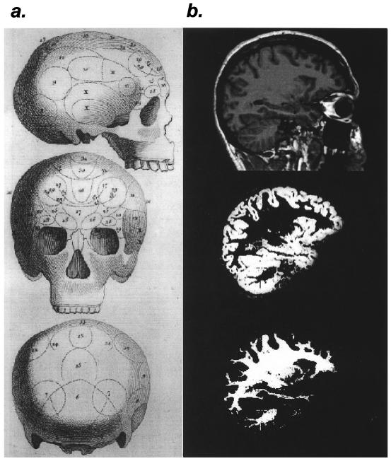
Structural studies of the brain, past and present. (a)
Phrenologist's map from the end of the 18th century. Bumps on the
skull were thought to reflect the size of the underlying brain.
[Reproduced with permission from John van Whye, The History of
Phrenology on the Web (http://www.jmvanwyhe.freeserve.co.uk ),
March 20, 2000. Originally published in The Philosophical
Magazine (1802), Vol. 14.] (b) Example of
voxel-based morphology as used in the Maguire et al. (1)
study. From top to bottom, T1-weighted anatomical image in a sagittal
plane containing the hippocampus, segmented gray matter from the same
image, and segmented white matter from the same image. [Images
courtesy of Timothy M. Ellmore, Laboratory of Brain and Cognition,
National Institute of Mental Health, Bethesda, MD.]
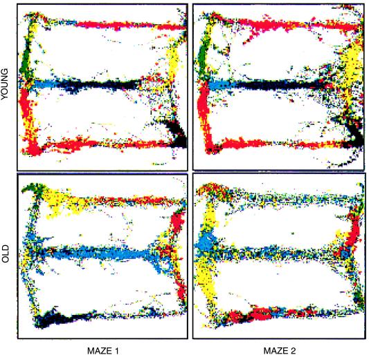
Multistability of place representation in old rats. Parallel recordings
of single units in hippocampal area CA1 made on two consecutive
experiences of a “Figure 8” maze (Maze 1, Maze 2; see figure 8 of
ref. 18) for one old and one young rat. Between each recording session,
the rats were removed from the room. Each color represents the spikes
of a single unit recorded as the rat traversed the maze. The rat's
movements through the maze are represented by the underlying gray
traces. For the young rat, the spatial representation is highly
consistent between visits. In contrast, the older rat exhibits an
almost complete redistribution of the firing fields between the two
visits. [Reproduced with permission from Barnes et al.
(1997) Nature (London) 388, 272–275
(Copyright 1997, Macmillan Magazines Ltd).]
Comment on
-
Navigation-related structural change in the hippocampi of taxi drivers.Proc Natl Acad Sci U S A. 2000 Apr 11;97(8):4398-403. doi: 10.1073/pnas.070039597. Proc Natl Acad Sci U S A. 2000. PMID: 10716738 Free PMC article.
References
-
- Witelson S F, Kigar D L, Harvey T. Lancet. 1999;353:2149–2153. - PubMed
-
- O'Keefe J, Nadel L. The Hippocampus as a Cognitive Map. Oxford: Clarendon; 1978.
-
- Squire L. Psychol Rev. 1992;99:195–231. - PubMed
-
- Cohen N J, Eichenbaum H. Memory, Amnesia, and the Hippocampal System. Cambridge, MA: MIT Press; 1993.
Publication types
MeSH terms
Grants and funding
LinkOut - more resources
Full Text Sources
