. 2000 Apr 25;97(9):4530-4.
doi: 10.1073/pnas.97.9.4530.

Divergence of larval morphology between Drosophila sechellia and its sibling species caused by cis-regulatory evolution of ovo/shaven-baby

Affiliations

Divergence of larval morphology between Drosophila sechellia and its sibling species caused by cis-regulatory evolution of ovo/shaven-baby

E Sucena et al. Proc Natl Acad Sci U S A. .

Abstract

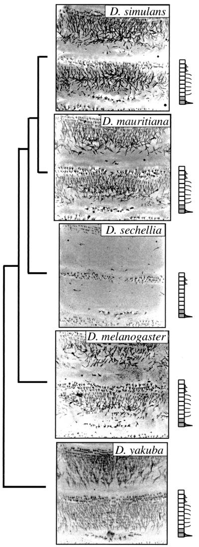
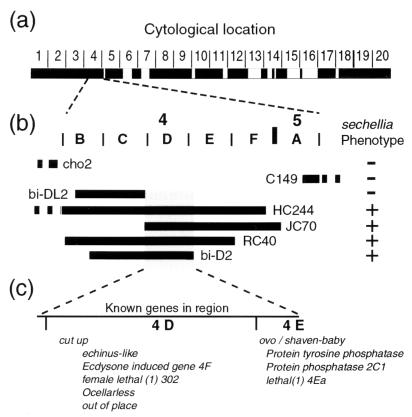
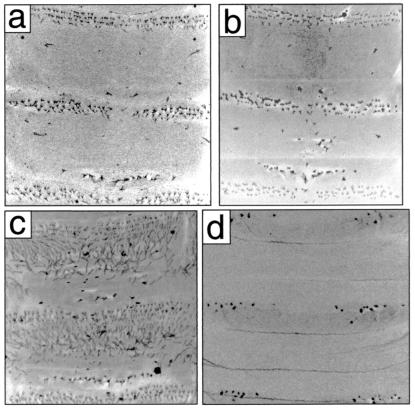
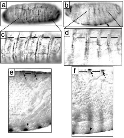
We report an extreme morphological difference between Drosophila sechellia and related species of the pattern of hairs on first-instar larvae. On the dorsum of most species, the posterior region of the anterior compartment of most segments is covered by a carpet of fine hairs. In D. sechellia, these hairs have been lost and replaced with naked cuticle. Genetic mapping experiments and interspecific complementation tests indicate that this difference is caused, in its entirety, by evolution at the ovo/shaven-baby locus. The pattern of expression of the ovo/shaven-baby transcript is correlated with this morphological change. The altered dorsal cuticle pattern is probably caused by evolution of the cis-regulatory region of ovo/shaven-baby in the D. sechellia lineage.


Figures

Figure 1
Figure 1
The phylogenetic distribution of dorsal hair patterns for five members of the D. melanogaster species group. A phylogeny of these species is shown (Left, modified from ref. 36). Confocal micrographs are shown for abdominal segments 1 and 2 for each species. A cartoon of the pattern of hairs is shown beside each micrograph. In the cartoons, cells in the anterior and posterior compartment of the segment are shown as white and gray rectangles, respectively. The relative positions of the three types of cuticular projections, short denticles, fine hairs, and large denticles are illustrated (see text). Anterior is up. The dorsal hair patterns for the remaining members of the group (Drosophila erecta, Drosophila orena, and Drosophila teissieri) are similar to Drosophila yakuba (not shown).
Figure 2
Figure 2
Localization of the evolved gene by failure of complementation of X chromosome deficiencies. (a) The cytological regions covered by deficiencies that produced viable larvae when crossed to D. sechellia males are shown as black boxes. Approximately 75% of the chromosome was screened successfully with deficiencies. In the original screen, only Df(1)JC70 produced larvae with the D. sechellia hair pattern in the expected ratio (n = 57).[Some of the crosses yielded single larvae with a D. sechellia hair pattern. These larvae are likely the result of meiotic nondisjunction in the female parent generating nullo X eggs fertilized by X-bearing D. sechellia sperm, which in turn generated XO embryos displaying the D. sechellia hair pattern. Nondisjunction is elevated in stocks carrying balancer chromosomes and stocks with XXY females (37)]. (b) Further localization to the 4C15-E1 cytological region was performed with overlapping deficiencies. Regions deleted by deficiencies are indicated by bold horizontal lines with the name of the deficiency next to the line. The continuation of a deficiency outside this region is shown by a dashed line. Deficiencies producing larvae with a D. sechellia hair pattern in the expected frequencies when crossed to D. sechellia males are indicated with a plus sign. Deficiencies producing only larvae with a hair pattern typical of D. melanogaster are indicated by a minus sign. The distal limit of the evolved gene is defined by the right breakpoint of Df(1)bi-DL2 (4C15-D1) and the left breakpoint of Df(1) JC70 (4C15–16), and the proximal limit is defined by the right breakpoint of Df(1)bi-D2 (4D7-E1). (c) Genes known to exist within this region are listed in their approximate cytological location. The genes cut up and ovo/svb have been localized previously to the small regions, 4D1–3 and 4E1, respectively, and the three genes Protein tyrosine phosphatase 4E, Protein phosphatase 2C1, and lethal(1)4Ea have been localized previously to 4E1–2. The remaining genes shown have been localized to large regions that include 4D-E1 (see http://flybase.bio.indiana.edu for details).
Figure 3
Figure 3
Confocal micrographs of the first and second abdominal segments of D. sechellia (a) are similar to those of a hybrid between a D. melanogaster svb1 mutant and D. sechellia (b). In contrast, a hybrid of wild-type D. melanogaster and D. sechellia (c) displays a cuticular pattern similar to D. melanogaster (see Fig. 1). The D. melanogaster svb1 mutations leads to complete loss of denticles and hairs on the dorsal surface (not shown) and loss of most denticles on the ventral surface (d). [In some svb1/D. sechellia larvae, small patches of hairs in the middle of naked cuticle were occasionally observed (b). Such hairs were never observed in hybrid backcrosses and in crosses between svb deficiencies and D. sechellia, suggesting that svb1 is not a complete loss-of-function allele.] Anterior is up.
Figure 4
Figure 4
The pattern of svb expression in D. melanogaster (a, c, and e) and D. sechellia (b, d, and f) stage 17 embryos. Dorsal views are shown in a, c, and d, dorso–lateral in b, and lateral optical cross sections in e and f. The svb transcript is detected in both species in rows corresponding to the dorsal stout denticles (a–d and arrows in e and f). svb transcript is detected at lower levels in the rows giving rise to fine hairs in D. melanogaster (horizontal bars in c and e) and is not detected in the corresponding positions in D. sechellia (horizontal bars in d and f). The cells differentiating the ventral denticle belts express high levels of svb (arrowheads in b, e, and f). Anterior is left in all images, and dorsal is up in b, e, and f.

References

    1. Averof M, Akam M. Nature (London) 1995;376:420–423. - PubMed
    1. Carroll S B, Weatherbee S D, Langeland J A. Nature (London) 1995;375:58–61. - PubMed
    1. Panganiban G, Sebring A, Nagy L, Carroll S. Science. 1995;270:1363–1366. - PubMed
    1. Gellon G, McGinnis W. BioEssays. 1998;20:116–125. - PubMed
    1. Fisher R A. The Genetical Theory of Natural Selection. NY: Dover; 1930. pp. 1–188.

Publication types

LinkOut - more resources