. 2000 Apr 25;97(9):4573-8.
doi: 10.1073/pnas.080508097.

The crystal structure of palmitoyl protein thioesterase 1 and the molecular basis of infantile neuronal ceroid lipofuscinosis

Affiliations

The crystal structure of palmitoyl protein thioesterase 1 and the molecular basis of infantile neuronal ceroid lipofuscinosis

J J Bellizzi 3rd et al. Proc Natl Acad Sci U S A. .

Abstract

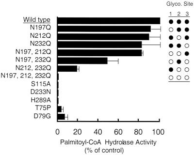
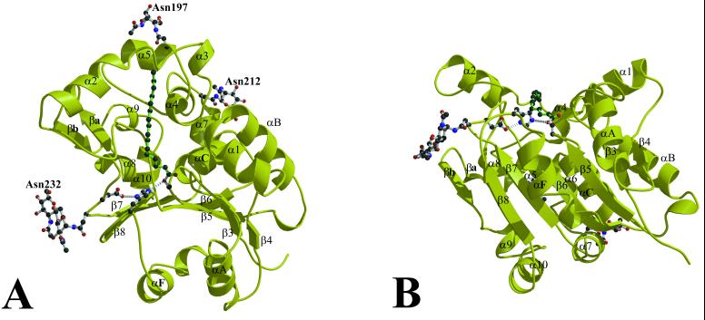
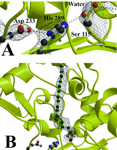
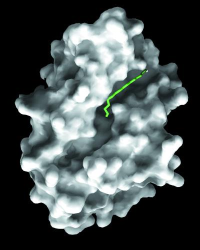
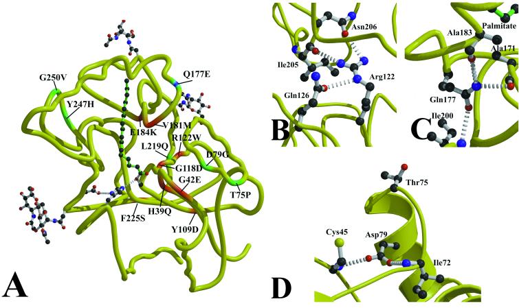
Mutations in palmitoyl-protein thioesterase 1 (PPT1), a lysosomal enzyme that removes fatty acyl groups from cysteine residues in modified proteins, cause the fatal inherited neurodegenerative disorder infantile neuronal ceroid lipofuscinosis. The accumulation of undigested substrates leads to the formation of neuronal storage bodies that are associated with the clinical symptoms. Less severe forms of PPT1 deficiency have been found recently that are caused by a distinct set of PPT1 mutations, some of which retain a small amount of thioesterase activity. We have determined the crystal structure of PPT1 with and without bound palmitate by using multiwavelength anomalous diffraction phasing. The structure reveals an alpha/beta-hydrolase fold with a catalytic triad composed of Ser115-His289-Asp233 and provides insights into the structural basis for the phenotypes associated with PPT1 mutations.


Figures

Figure 1
Figure 1
Palmitoyl-CoA hydrolase activity in COS cell lysates expressing wild-type and mutant human PPT1s. Activity is expressed as a percentage of wild-type activity over control cells transfected with vector alone. Each value represents the mean ± SE from three or more independent experiments. As confirmed by the crystal structure, Ser115, Asp233, and His289 are essential for activity and comprise the catalytic triad of PPT1. To assess the effect of glycosylation site mutations on PPT1 activity, the three asparagine glycosylation sites in PPT1 were mutated singly or in combination as shown. Filled circles indicate glycosylation site occupancy. Two mutations associated with juvenile onset NCL, Thr75Pro and Asp79Gly, are shown to exhibit detectable residual PPT activity.
Figure 2
Figure 2
The crystal structure of PPT1 complexed with palmitate. β3–β8 and αA, αB, αC, and αF are elements of the canonical α/β hydrolase fold and are labeled according to the nomenclature of Ollis (27). The palmitate, the catalytic triad, and the glycosylated asparagines are indicated. B is a rotation by 90° from A. Figs. 2, 3, and 5 were prepared by using bobscript (28), molscript (29), and raster3d (30).
Figure 3
Figure 3
Active site of PPT1. (A) The active site of native PPT1. A water molecule occupies the oxyanion hole, hydrogen-bonded to Ser115, Met41, and Gln116. The solvent-flattened multiwavelength anomalous diffraction electron density with experimental phases is superimposed on the model, contoured at 1.4σ. (B) The active site of the PPT1/palmitate complex. A simulated annealing Fo-Fc map, generated by using cns (24) with Ser115 and palmitate omitted from the calculation, is shown contoured at 1.5σ.
Figure 4
Figure 4
The palmitate binding groove. The molecular surface was calculated by using grasp (31). The palmitate (green stick model) occupies a deep, narrow groove on the hydrophobic face of the protein.
Figure 5
Figure 5
NCL mutations in PPT1. (A) Sites of clinical NCL mutations in PPT1 are mapped onto the peptide backbone. INCL mutations are displayed in red, a mutation causing LINCL symptoms is in blue, and JNCL mutations are in green. (B) The most common INCL mutation (Arg122Trp) leads to the loss of three hydrogen bonds and a steric and polarity mismatch with the surrounding residues, resulting in misfolded protein. (C) The Gln177Glu mutation is predicted to cause the loss of hydrogen bonds to Ala171 and Ala183, two residues that contact palmitate. This mutation results in a less severe phenotype that is clinically indistinguishable from classical LINCL. (D) Two mutations on α1 lead to JNCL. Trp75Pro may alter the beginning of α1 due to the conformational restraints on Pro, and Asp79Glu loses hydrogen bonds to Cys45 and Ile72.

References

    1. Santavuori P, Haltia M, Rapola J. Dev Med Child Neurol. 1974;16:644–653. - PubMed
    1. Vesa J, Hellsten E, Verkruyse L A, Camp L A, Rapola J, Santavuori P, Hofmann S L, Peltonen L. Nature (London) 1995;376:584–587. - PubMed
    1. Hellsten E, Vesa J, Speer M C, Makela T P, Jarvela I, Alitalo K, Ott J, Peltonen L. Genomics. 1993;16:720–725. - PubMed
    1. Hofmann S L, Das A K, Yi W, Lu J-Y, Wisniewski K E. Mol Genet Metab. 1999;66:234–239. - PubMed
    1. Verkruyse L A, Hofmann S L. J Biol Chem. 1996;271:15831–15836. - PubMed

Publication types

Substances

LinkOut - more resources