. 2000 Apr 25;97(9):4666-71.
doi: 10.1073/pnas.090082297.

Outer mitochondrial membrane permeability can regulate coupled respiration and cell survival

Affiliations

Outer mitochondrial membrane permeability can regulate coupled respiration and cell survival

M G Vander Heiden et al. Proc Natl Acad Sci U S A. .

Abstract

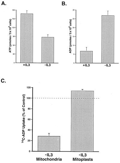
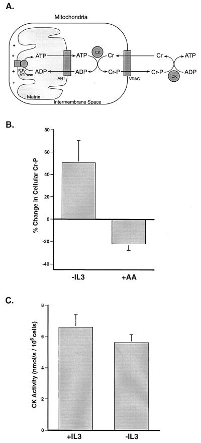
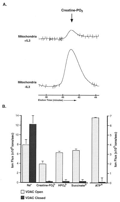
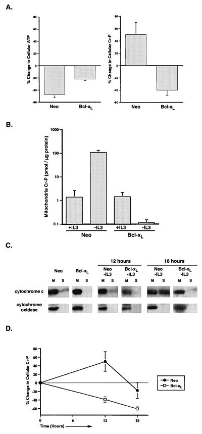
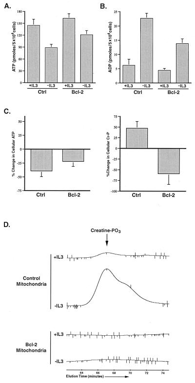
Coupled cellular respiration requires that ATP and ADP be efficiently exchanged between the cytosol and the mitochondrial matrix. When growth factors are withdrawn from dependent cells, metabolism is disrupted by a defect in ATP/ADP exchange across the mitochondrial membranes. Unexpectedly, we find that this defect results from loss of outer mitochondrial membrane permeability to metabolic anions. This decrease in anion permeability correlates with the changes in conductance properties that accompany closure of the voltage-dependent anion channel (also known as mitochondrial porin). Loss of outer membrane permeability (i) results in the accumulation of stored metabolic energy within the intermembrane space in the form of creatine phosphate, (ii) is prevented by the outer mitochondrial membrane proteins Bcl-x(L) and Bcl-2, and (iii) can be reversed by growth factor readdition. If outer membrane impermeability persists, the disruption of mitochondrial homeostasis culminates in loss of outer mitochondrial membrane integrity, cytochrome c redistribution, and apoptosis. The recognition that outer membrane permeability is regulated under physiological conditions has important implications for the understanding of bioenergetics and cell survival.


Figures

Figure 1
Figure 1
Growth factor withdrawal results in a mitochondrial ATP/ADP exchange defect that is corrected by removal of the outer mitochondrial membrane. (A) IL-3-dependent FL5.12 cells were cultured in the presence (+IL3) or absence (−IL3) of IL-3 for 12 h before the determination of cellular ATP. The data shown are the mean (+ one SEM) of four independent experiments. The difference in ATP was statistically significant by Student's t test (P < 0.01). (B) Cells were cultured in the presence (+IL3) or absence (−IL3) of IL-3 for 12 h, and the amount of cellular ADP present was determined. The data shown are the mean (+SEM) of four independent measurements. The difference in ADP was statistically significant by Student's t test (P < 0.01). (C) Mitochondria were isolated from cells cultured in the presence or absence of IL-3 for 12 h. Where indicated, the outer mitochondrial membrane was removed by using digitonin (mitoplasts). The resultant mitoplasts contained less than 1% of the intermembrane space molecule phosphocreatine found in mitochondria yet retained the ability to carry out the ANT-specific import of ADP. ANT-dependent ADP uptake of mitochondria and mitoplasts was assessed by comparing the uptake of 14C-ADP in the presence or absence of atractyloside, a specific inhibitor of the ANT. The mean ANT-dependent uptake for mitochondria and mitoplasts from cells deprived of IL-3 (−IL3) is shown as a percentage of that observed in mitochondria and mitoplasts from cells growing in the presence of IL-3 (+SEM). The difference in ADP uptake between mitochondria and mitoplasts was statistically significant by Student's t test (P < 0.01).
Figure 2
Figure 2
Growth factor-deprived cells accumulate creatine phosphate. (A) A schematic representation of the shuttle mechanism through which creatine/creatine phosphate (Cr/Cr-P) can buffer cytosolic ATP levels. (B) Cells were withdrawn from IL-3 for 12 h or treated for 30 min in the presence of IL-3 with the electron transport inhibitor, antimycin A, before the preparation of perchloric acid extracts. The amount of phosphocreatine present in the extracts was determined by using HPLC. The percent change in cellular phosphocreatine measured after IL-3 withdrawal (−IL3) and the addition of antimycin A (+AA) is shown. The data presented are the mean (+SEM) of at least four independent experiments. The changes in phosphocreatine observed in both cases were statistically significant by Student's t test (P < 0.05). (C) Creatine kinase (CK) catalyzes the reaction: ADP + Cr-P ↔ ATP + Cr, hence creatine kinase activity was assessed by measuring the ability of saponin-lysed cells to convert creatine phosphate to creatine in the presence of ADP. At the low concentrations used, saponin preferentially disrupts the plasma membrane of cells while leaving intracellular membranes intact. The CK activity present in lysates prepared from equal numbers of cells cultured in the presence (+IL3) or absence (−IL3) of IL-3 for 12 h is depicted. Data shown are the mean (+ one SD) of three independent measurements.
Figure 3
Figure 3
Anion flux through the outer mitochondrial membrane and through isolated VDAC channels can be regulated. (A) Mitochondria were isolated from cells cultured in the presence (+IL3) or absence (−IL3) of IL-3 for 12 h, and perchloric acid extracts normalized to mitochondrial protein were prepared. The amount of creatine phosphate (creatine–PO3) present in the mitochondrial extracts was determined by HPLC. The peak corresponding to the amount of creatine–PO3 present in the mitochondria under each condition is shown. The data presented are representative of results from four independent experiments, and the increase observed in all cases was statistically significant by Student's t test (P < 0.02). The creatine-PO3 peak of both samples was confirmed by coinjection of purified creatine–PO3 (not shown). (B) The net ion flux through single VDAC channels in the high-conducting (open) state and in the low-conducting (closed) state. All fluxes shown are the mean (+SEM) and reflect ion flow in the absence of an electrical potential and in the presence of a concentration difference of 100 mM salt. The difference in phosphocreatine flux through VDAC in the open and closed states was statistically significant by Student's t test (P < 0.01).
Figure 4
Figure 4
Loss of outer mitochondrial membrane permeability after growth factor withdrawal is prevented by Bcl-xL expression. (A) Bcl-xL- and control- (Neo) transfected FL5.12 cells were cultured in the presence or absence of IL-3 for 12 h before the determination of ATP and phosphocreatine. The amount of ATP was determined by the luciferin/luciferase method, and the amount of phosphocreatine was determined by HPLC. The percent change in cellular ATP and creatine phosphate (Cr-P) measured on IL-3 withdrawal for each population is shown. The data presented are the mean (+SEM) of at least four independent determinations. The changes in ATP and phosphocreatine observed in both cases were statistically significant by Student's t test (P < 0.01). (B) Bcl-xL- and control- (Neo) transfected cells were cultured in the presence or absence of IL-3 for 12 h, and perchloric acid extracts normalized to mitochondrial protein were prepared. The mean (+SEM) amount of creatine phosphate present in the mitochondrial extracts from four independent experiments is shown. (C) Bcl-xL- and control- (Neo) transfected cells that were cultured in the presence of IL-3 or deprived of IL-3 (-IL3) for the time indicated were mechanically lysed and separated into mitochondrial (M) and S-100 (S) fractions. The cytosol is represented in the S-100 fraction. The amount of cytochrome c and cytochrome oxidase subunit IV in each fraction was determined by Western blot analysis. (D) Perchloric acid extracts were prepared from equal numbers of Bcl-xL- and control- (Neo) transfected cells that were withdrawn from IL- 3 for the period indicated. Cellular phosphocreatine was measured by using HPLC, and the percent change in phosphocreatine over time is graphed. The data presented are the mean (+SEM) of five independent experiments.
Figure 5
Figure 5
Growth factor withdrawal in Baf3 cells results in mitochondrial creatine phosphate accumulation that is prevented by Bcl-2 expression. (A) Control (Ctrl) and Bcl-2-transfected Baf3 cells were cultured in the presence (+IL3) or absence (−IL3) of IL-3 for 12 h before the determination of cellular ATP. The data shown are the mean (+SEM) of four independent experiments. The fall in ATP observed on IL-3 withdrawal was statistically significant by Student's t test (P < 0.05). (B) Control (Ctrl) and Bcl-2-transfected cells were cultured in the presence (+IL3) or absence (−IL3) of IL-3 for 12 h, and the amount of cellular ADP present was determined. The data shown are the mean (+SEM) of four independent measurements. The increase in ADP on IL-3 withdrawal was statistically significant by Student's t test (P < 0.05). (C) Control and Bcl-2-transfected cells were cultured in the presence or absence of IL-3 for 12 h before the determination of ATP and phosphocreatine. The percent change in cellular ATP and creatine phosphate (Cr-P) measured on IL-3 withdrawal for each population is shown. The data presented are the mean (+SEM) of four independent determinations. The changes in ATP and phosphocreatine observed in both cases were statistically significant by Student's t test (P < 0.05). (D) Control and Bcl-2-transfected cells were cultured in the presence or absence of IL-3 for 12 h, and perchloric acid extracts normalized to mitochondrial protein were prepared. The amount of creatine phosphate (creatine-PO3) present in the mitochondrial extracts was determined by HPLC. The peak corresponding to the amount of creatine-PO3 present in the mitochondria under each condition is shown. The creatine-PO3 peak in the samples was confirmed by coinjection of purified creatine-PO3 (not shown).

References

    1. Reed J C. Cell. 1997;91:559–562. - PubMed
    1. Vander Heiden M G, Chandel N S, Williamson E K, Schumacker P T, Thompson C B. Cell. 1997;91:627–637. - PubMed
    1. Vander Heiden M G, Chandel N S, Schumacker P T, Thompson C B. Mol Cell. 1999;3:159–167. - PubMed
    1. Wallace D C. Science. 1999;283:1482–1488. - PubMed
    1. Rostovtseva T, Colombini M. Biophys J. 1997;72:1954–1962. - PMC - PubMed

Publication types