. 2000 Apr 25;97(9):4914-9.
doi: 10.1073/pnas.090092797.

Colocalization and coassembly of two human brain M-type potassium channel subunits that are mutated in epilepsy

Affiliations

Colocalization and coassembly of two human brain M-type potassium channel subunits that are mutated in epilepsy

E C Cooper et al. Proc Natl Acad Sci U S A. .

Abstract

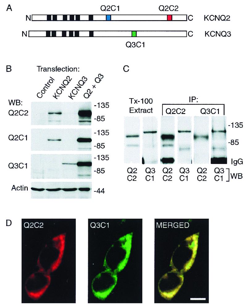
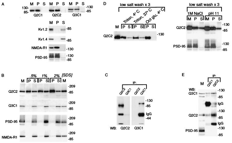
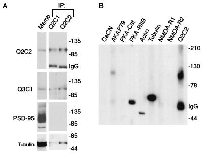
Acetylcholine excites many central and autonomic neurons through inhibition of M-channels, slowly activating, noninactivating voltage-gated potassium channels. We here provide information regarding the in vivo distribution and biochemical characteristics of human brain KCNQ2 and KCNQ3, two channel subunits that form M-channels when expressed in vitro, and, when mutated, cause the dominantly inherited epileptic syndrome, benign neonatal familial convulsions. KCNQ2 and KCNQ3 proteins are colocalized in a somatodendritic pattern on pyramidal and polymorphic neurons in the human cortex and hippocampus. Immunoreactivity for KCNQ2, but not KCNQ3, is also prominent in some terminal fields, suggesting a presynaptic role for a distinct subgroup of M-channels in the regulation of action potential propagation and neurotransmitter release. KCNQ2 and KCNQ3 can be coimmunoprecipitated from brain lysates. Further, KCNQ2 and KCNQ3 are coassociated with tubulin and protein kinase A within a Triton X-100-insoluble protein complex. This complex is not associated with low-density membrane rafts or with N-methyl-d-aspartate receptors, PSD-95 scaffolding proteins, or other potassium channels tested. Our studies thus provide a view of a signaling complex that may be important for cognitive function as well as epilepsy. Analysis of this complex may shed light on the unknown transduction pathway linking muscarinic acetylcholine receptor activation to M-channel inhibition.


Figures

Figure 1
Figure 1
Characterization of heterologously expressed KCNQ2 and KCNQ3 and subunit-specific antibodies. (A) Diagram of the KCNQ2 and KCNQ3 polypeptides predicted by cDNA sequences, showing the position of the six putative membrane-spanning segments (black boxes), and the three segments within the C-terminal domains corresponding to synthetic peptides used for raising subunit-specific antibodies Q2C1, Q2C2, and Q3C1. (B) Immunoblots of whole-cell lysates prepared from HEK293 cells transfected with KCNQ2 alone, KCNQ3 alone, or cotransfected with both subunits. (C) Solubilization and coimmunoprecipitation of KCNQ2 and KCNQ3 expressed in HEK293 cells. Cells cotransfected with KCNQ2 and KCNQ3 were lysed under nondenaturing conditions with 1% Triton X-100. The lysates were cleared of nuclei and insoluble material by centrifugation, then either reserved (Tx-100 extract) or incubated with Q2C2 or Q3C1 antibodies. Whole-cell extracts and immunoprecipitates were probed with Q2C2, then stripped and reprobed with Q3C1. (D) Immunofluorescence staining of cotransfected HEK cells shows predominantly colocalized pattern of expression of KCNQ2 and KCNQ3. (Scale bar equals 10 μm.)
Figure 2
Figure 2
Immunolocalization of KCNQ2 and KCNQ3 in human cortex and hippocampus. (A) Low-power view (bar equals 1 mm) of transverse section through human hippocampal formation after immunoperoxidase staining using Q2C2 antibodies and light counterstaining with hematoxylin. Subregions indicated are the dentate gyrus (DG), sections CA3 and CA1 of the hippocampus, and the subiculum (sub). Regions shown in red boxes represent locations shown at higher power in E–H. (B) Control experiment showing immunostaining was abolished by preincubation of antibodies with antigenic peptide. Adjoining sections were incubated with Q2C2 antibodies in the presence (Left) or absence (Right) of excess Q2C2 peptide. (Left) Only purple–blue counterstain of neurons and glia. (Right) Brown immunoperoxidase staining of neuronal somata, proximal dendrites, and lighter neuropil staining. (C) Confocal images of Q2C2 (Left) and Q3C1 (Right) staining of temporal cortex (parahippocampal gyrus) neurons. Red shows channel antibody staining in a punctate somotodendritic distribution; green shows brain autofluorescence, mostly associated with lipofuscin in perinuclear vesicular structures. (Scale bar equals 10 μm.) (D) Q2C2 staining of mossy fiber bundles in the dentate hilar region. (E) Q2C2 (Left) and Q3C1 (Right) staining of polymorphic neurons in the dentate hilar region. (Arrows indicate stained neuronal somata; asterisks indicate neuropil, which is stained more heavily with Q2C2 than Q3C1.) (F) Q2C2 (Left) and Q3C1 (Right) staining of neurons in the CA3 pyramidal cell layer. (G) Q2C2 (Left) and Q3C1 (Right) staining of pyramidal cells in the subiculum. (H) Staining of the neuropil in the dentate hilar and inner molecular layers by Q2C2 but not Q3C1. Arrows indicate hilar neuronal somata, labeled by both antibodies; brackets indicate inner molecular layer staining by Q2C2 but not Q3C1. (Scale bar equals 100 μm.) Pm, polymorphic layer; gc, granule cell layer; iml, inner (associational-commissural) molecular layer; oml, outer (perforant path) molecular layer. (Scale bar in B equals 20 μm in B, D–G.)
Figure 3
Figure 3
Heteromeric human brain KCNQ2/KCNQ3 channels are contained within a Triton X-100-resistant subcellular fraction. (A) Insolubility of KCNQ2 and KCNQ3. Human cortical membranes were solubilized in 1% Triton X-100, and soluble and insoluble fractions were separated by centrifugation. Starting membranes (M) and insoluble (P, pellet) and soluble (S) fractions were probed with antibodies against KCNQ subunits, Shaker family potassium channel subunits (Kv1.2, Kvl.4), a glutamate receptor subunit (NMDA-R1), and PSD-95. The Shaker subunits were efficiently solubilized by Triton X-100; the KCNQ subunits, NMDA-R1 and PSD-95 were poorly solubilized. (B) Solubilization by SDS at 4°C. Human brain membranes were solubilized with SDS at the indicated concentrations on ice as described in Materials and Methods, and soluble and insoluble fractions were separated by centrifugation. KCNQ3, NMDA-R1, and PSD-95 controls were efficiently solubilized by cold SDS, but the majority of KCNQ2 remained associated with the insoluble fraction. (C) SDS-solubilized KCNQ2 and KCNQ3 are not coassociated. After solubilization of brain membranes using 1% SDS/4°C, Q2C1 and Q3C2 immunoprecipitates were probed with the indicated antibodies. (D) Solubilization of KCNQ2/KCNQ3 complexes (but not PSD-95) by Triton X-100 after low salt washes, high salt, and pH 11. Membranes were washed as described in Materials and Methods, then solubilized as indicated with octyl glucoside or Triton X-100 at 4°C or 37°C as indicated. KCNQ2 and KCNQ3 (not shown) were partly solubilized by Triton X-100 after low-salt washes, sequential low/high-salt washes, or pH 11, but not by octyl glucoside, and solubilization with Triton X-100 at 37°C after low-salt washes was not more effective than at 4°C. PSD-95 remained in pellet after salt or pH 11 treatment. (E) Coimmunoprecipitation of KCNQ2 and KCNQ3 by either Q2C2 or Q3C1 antibodies. Membranes were low-salt washed three times, extracted at pH 11, solubilized with Triton X-100, and cleared of insoluble material by centrifugation. KCNQ2 and KCNQ3 (but not PSD-95) were both immunoprecipitated by antibodies specific for either subunit.
Figure 4
Figure 4
KCNQ3, cytoskeletal proteins, and PKA are associated with KCNQ2 in a Triton X-100 insoluble complex. (A) KCNQ2-associated proteins were affinity-purified using Q2C1 or Q2C2 antibodies, and probed with the indicated antibodies. KCNQ2, KCNQ3, and tubulin were detected, but PSD-95, although abundant in the starting membranes, was absent in the affinity-purified fraction. (B) AKAP79, PKA RIIB, tubulin, and actin are present in the Q2C2 affinity-purified complex, but calcineurin, CaM Kinase, NMDA-R1, and NMDA-R2 are not present.

References

    1. Ptacek L. Am J Med. 1998;105:58–70. - PubMed
    1. Noebels J L. Neuron. 1996;16:241–244. - PubMed
    1. Cooper E C, Jan L Y. Proc Natl Acad Sci USA. 1999;96:4759–4766. - PMC - PubMed
    1. Leppert M, Anderson V E, Quattlebaum T, Stauffer D, O'Connell P, Nakamura Y, Lalouel J M, White R. Nature (London) 1989;337:647–648. - PubMed
    1. Singh N A, Charlier C, Stauffer D, DuPont B R, Leach R J, Melis R, Ronen G M, Bjerre I, Quattlebaum T, Murphy J V, et al. Nat Genet. 1998;18:25–29. - PubMed

Publication types

MeSH terms