. 2000 Apr 25;97(9):4979-84.
doi: 10.1073/pnas.97.9.4979.

Conserved plant genes with similarity to mammalian de novo DNA methyltransferases

Affiliations

Conserved plant genes with similarity to mammalian de novo DNA methyltransferases

X Cao et al. Proc Natl Acad Sci U S A. .

Abstract

DNA methylation plays a critical role in controlling states of gene activity in most eukaryotic organisms, and it is essential for proper growth and development. Patterns of methylation are established by de novo methyltransferases and maintained by maintenance methyltransferase activities. The Dnmt3 family of de novo DNA methyltransferases has recently been characterized in animals. Here we describe DNA methyltransferase genes from both Arabidopsis and maize that show a high level of sequence similarity to Dnmt3, suggesting that they encode plant de novo methyltransferases. Relative to all known eukaryotic methyltransferases, these plant proteins contain a novel arrangement of the motifs required for DNA methyltransferase catalytic activity. The N termini of these methyltransferases contain a series of ubiquitin-associated (UBA) domains. UBA domains are found in several ubiquitin pathway proteins and in DNA repair enzymes such as Rad23, and they may be involved in ubiquitin binding. The presence of UBA domains provides a possible link between DNA methylation and ubiquitin/proteasome pathways.


Figures
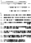
Figure 1
Figure 1
(A) Schematic diagram of the domain structures of Dnmt3b, DRM2, and Zmet3. Figure is drawn to scale. Shaded boxes show the different motifs present in these proteins, including the PWWP and cysteine rich (C-rich) motifs present in Dnmt3b and the UBA domains present in DRM2 and Zmet3. Roman numerals denote the motifs of the methyltransferase catalytic domains. (B) Alignment of DRM2 of Arabidopsis thaliana with Zmet3 of Zea mays and the methyltransferase catalytic domains of mouse Dnmt3b (GenBank accession no. AF068628) and Danio rerio Zmt3 (Danmt3; accession no. AF135438). Alignments were done in clustalX 1.8 using default parameters and shaded in MacBoxShade 1.0.8 (Michael D. Baron, Institute for Animal Health, Surrey, U.K.). Identical residues are shown with a black background and similar residues with a gray background. Dashes show putative nuclear localization sequences that are conserved in the plant proteins. Pound symbols (#) show the point of rearrangement of the plant proteins relative to the animal proteins. Here the numbering of the animal methyltransferases begins at amino acid 581 for Dnmt3b and 558 for Danmt3. Conserved catalytic motifs I–VI and IX–X are marked. Asterisks denote conserved amino acids present in each motif (11). Brackets show the exact region used in the alignments that were used to produce the tree shown in Fig. 3.
Figure 2
Figure 2
(A) An alignment of motifs X and I of HhaI with the sequence of DRM2 spanning the point of motif rearrangement. HhaI residues underlined are the last residue of motif X and the first residue of motif I and correspond the positions in B marked by arrows. Alignments were done in clustalX and shaded in boxshade. (B) rasmol-generated image (Roger Sayle, Glaxo Research and Development, Greenford, Middlesex, U.K.) of Protein Database ID 5MHT (ternary structure of HhaI methyltransferase with hemimethylated DNA and S-adenosylhomocysteine) (36, 37). DNA helix is shown in black, the majority of HhaI is shaded gray, motif I is red, and motif X is blue. The colored regions correspond to those shown in the alignment in A, which are residues 9–32 for motif I and residues 299–322 for motif X. Arrows shows the last residue of motif X and first residue of motif I.
Figure 3
Figure 3
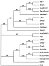
Phylogenetic relationships of DNA methyltransferases. Tree shows nodes with >50% bootstrap support. Abbreviations and accession numbers, in parentheses, are as follows: MET1, Arabidopsis thaliana maintenance methyltransferase MET1 (P34881); Zmet1, Zea mays MET1-like protein (AF0063403); MusDnmt1, mouse maintenance methyltransferase Dnmt1 (P13864); DanioDnmt1, zebrafish Dnmt-like protein (AF097875); AscMasc2, Ascobolus immersus Masc2 (AF030976); CMT1, Arabidopsis thaliana chromomethylase 1 (AF039372); Zmet2, Zea mays CMT-like protein (AF243043); CMT2, Arabidopsis thaliana chromomethylase 2 (AL021711); AscMasc1, Ascobolus immersus Masc1 (AF025475); BssHII, Bacillus stearothermophilus BssHII methylase (AF020002); MusDNMT2, mouse DNMT2 (AF045889); PMT, Schizosaccharomyces pombe PMTp1 (P40999); DRM1, Arabidopsis thaliana DRM1 (B62154); DRM2, Arabidopsis thaliana DRM2; Zmet3, Zea mays Zmet3; SoyDRM, soybean EST clone with similarity to DRM and Zmet3 (A1736568); MusDnmt3A, mouse de novo methyltransferase Dnmt3a (AF068625); MusDnmt3B, mouse de novo methyltransferase Dnmt3b (AF068628); DanioDnmt3, zebra fish Dnmt3-like protein (AF135438); MSPR, Bacillus subtilis bacteriophage SPR methyltransferase (P00476); and HhaI, bacterial (Haemophilus haemolyticus) HhaI methyltransferase used as an outgroup (P05102).
Figure 4
Figure 4
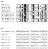
(A) clustalX alignment of the UBA domains predicted by PFAM (http://pfam.wustl.edu/) of DRM2 and Zmet3 with those of human p62 (accession no. U46751), yeast Rad23 (accession no. S66117), human Cbl (accession no. A43817), human ubiquitin C-terminal hydrolase 5 (accession no. P45974), Drosophila melanogaster ubiquitin-conjugating enzyme E2 (accession no. P52486), and human HHR23A (accession no. S44443). Below the alignment is the known structure of the second UBA domain of HHR23A, showing the three α-helices (- - -) (41) (Thomas Mueller and Juli Feigon, personal communication). *, Residues making important contributions to the hydrophobic core. #, Phe residue connecting the loop between helices α1 and α2 to the C terminus of helix α3. (B) Results of nearest-neighbor secondary structure prediction (NNSSP) on the Baylor College of Medicine Protein Web server (http://dot.imgen.bcm.tmc.edu:9331/pssprediction/pssp.html) (54), which predicts the presence of α-helices (a) or β-sheets (b). Gaps were introduced in the sequences as in the alignments in A.
Figure 5
Figure 5
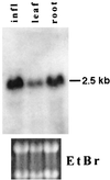
RNA blot analysis showing the size and abundance of DRM2 in different tissues. RNA from roots, leaves, or inflorescences was blotted and hybridized to a full-length DRM2 clone. Ethidium bromide staining of the ribosomal RNA bands is shown below as a loading control.

Similar articles

Cited by

References

    1. Martienssen R A, Richards E J. Curr Opin Genet Dev. 1995;5:234–242. - PubMed
    1. Bender J, Fink G R. Cell. 1995;83:725–734. - PubMed
    1. Jacobsen S E, Meyerowitz E M. Science. 1997;277:1100–1103. - PubMed
    1. Matzke M A, Matzke A J. Cell Mol Life Sci. 1998;54:94–103. - PMC - PubMed
    1. Finnegan E J, Peacock W J, Dennis E S. Proc Natl Acad Sci USA. 1996;93:8449–8454. - PMC - PubMed

Publication types

MeSH terms

Associated data