

Risk factors for proliferative vitreoretinopathy after primary vitrectomy: a prospective study
- PMID: 10781515
- PMCID: PMC1723478
- DOI: 10.1136/bjo.84.5.506
Risk factors for proliferative vitreoretinopathy after primary vitrectomy: a prospective study
Abstract
Aim: To assess clinical variables and vitreous protein as risk factors for the development of postoperative proliferative vitreoretinopathy (PVR).
Methods: A prospective study was conducted on 140 patients with a rhegmatogenous retinal detachment in whom a primary vitrectomy was performed. 12 clinical variables were recorded and vitreous samples obtained for measurement of protein concentration. Univariate and multivariate logistic regression analysis was used to determine the risk factors for PVR.
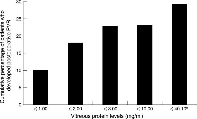
Results: Complete data were available for 136 of 140 patients. 40 of the 136 patients (29.4%) developed postoperative PVR. Univariate regression revealed that significant (p<0.05) risk factors included aphakia, presence of preoperative PVR, size of detachment, the use of silicone oil, and high vitreous protein level. Multivariate regression analysis revealed only aphakia (odds ratio 2.72), the presence of preoperative PVR (odds ratio 3.01), and high vitreous protein concentration (odds ratio 1.11) to be significant (p<0.05) independent, predictive risk factors for the development of PVR.
Conclusions: This study has shown that the significant risk factors for PVR are preoperative PVR, aphakia, and high vitreous protein levels. Two models (clinical factors only and clinical factors and vitreous protein) were constructed to predict the probability of developing postoperative PVR and may be used to identify those at risk for possible intravitreal pharmacological treatment.
Figures
References
Publication types
MeSH terms
Substances
LinkOut - more resources
Full Text Sources
Research Materials