. 2000 May;182(10):2761-70.
doi: 10.1128/JB.182.10.2761-2770.2000.

Enzymology of type IV macromolecule secretion systems: the conjugative transfer regions of plasmids RP4 and R388 and the cag pathogenicity island of Helicobacter pylori encode structurally and functionally related nucleoside triphosphate hydrolases

Affiliations

Enzymology of type IV macromolecule secretion systems: the conjugative transfer regions of plasmids RP4 and R388 and the cag pathogenicity island of Helicobacter pylori encode structurally and functionally related nucleoside triphosphate hydrolases

S Krause et al. J Bacteriol. 2000 May.

Abstract

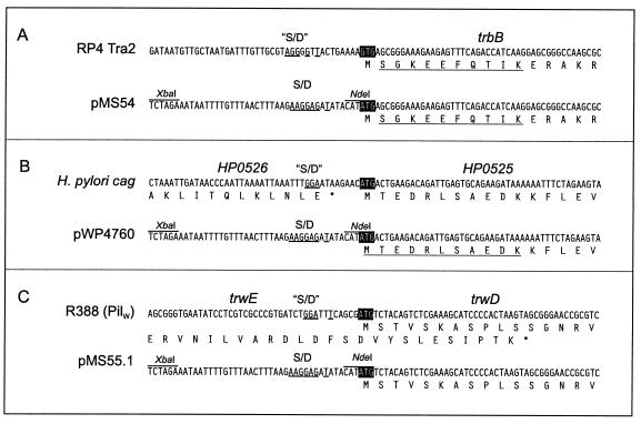
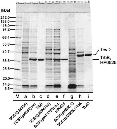
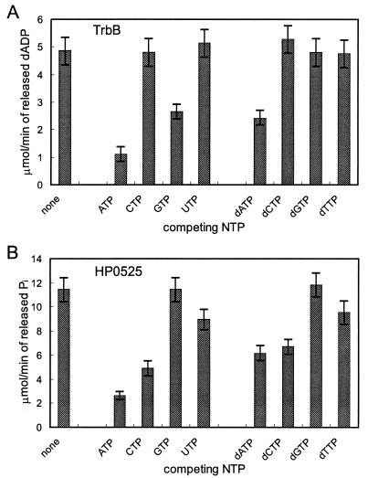
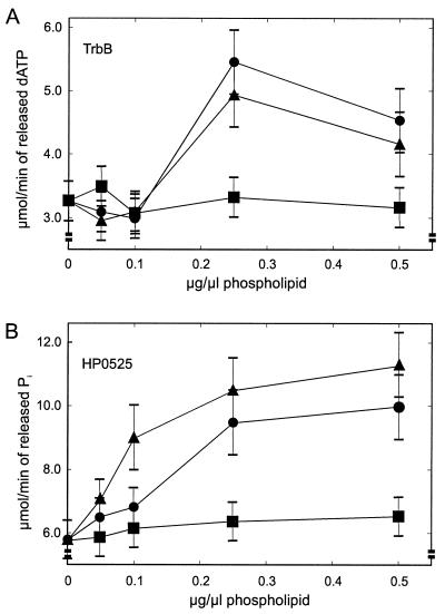
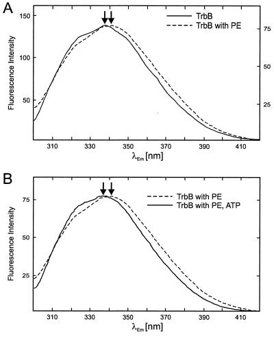
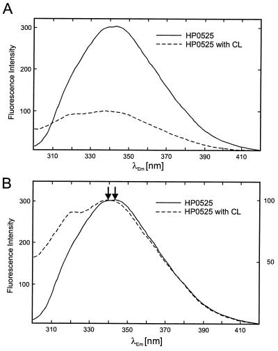
Type IV secretion systems direct transport of protein or nucleoprotein complexes across the cell envelopes of prokaryotic donor and eukaryotic or prokaryotic recipient cells. The process is mediated by a membrane-spanning multiprotein assembly. Potential NTPases belonging to the VirB11 family are an essential part of the membrane-spanning complex. Three representatives of these NTPases originating from the conjugative transfer regions of plasmids RP4 (TrbB) and R388 (TrwD) and from the cag pathogenicity island of Helicobacter pylori (HP0525) were overproduced and purified in native form. The proteins display NTPase activity with distinct substrate specificities in vitro. TrbB shows its highest specific hydrolase activity with dATP, and the preferred substrate for HP0525 is ATP. Analysis of defined TrbB mutations altered in motifs conserved within the VirB11 protein family shows that there is a correlation between the loss or reduction of NTPase activity and transfer frequency. Tryptophan fluorescence spectroscopy of TrbB and HP0525 suggests that both interact with phospholipid membranes, changing their conformation. NTPase activity of both proteins was stimulated by the addition of certain phospholipids. According to our results, Virb11-like proteins seem to most likely be involved in the assembly of the membrane-spanning multiprotein complex.


Figures

FIG. 1
FIG. 1
Construction of overexpression plasmids for RP4 trbB, cag HP0525, and R388 trwD. Upper panels, nucleotide sequences flanking the initiation codons of trbB (A), HP0525 (B), and trwD (C) in their native environments. Nucleotide sequences resembling Shine-Dalgarno sequences (“S/D”) are underlined. Initiation codons are marked by black boxes. Partial amino acid sequences of gene products are shown below the nucleotide sequences. Lower panels, genes were placed under translational control by the T7 gene 10 ribosomal binding site (S/D) by insertion into the expression vector pMS470Δ8. Underlined parts of the amino acid sequences were confirmed by N-terminal sequencing.
FIG. 2
FIG. 2
Purification of NTPases of type IV secretion pathways. Lane a, SCS1 (pMS54), no IPTG added; lane b, SCS1(pMS54), cells were induced for 5 h with 1 mM IPTG; lane c, TrbB, fraction V, 5 μg; lane d, SCS1(pWP4760), no IPTG added; lane e, SCS1(pWP4760), cells were induced for 5 h with 1 mM IPTG; lane f, HP0525, fraction IV, 5 μg; lane g, SCS1(pMS55.1), no IPTG added; lane h, SCS1(pMS55.1) cells were induced for 5 h with 1 mM IPTG; lane i, TrwD, fraction V, 5 μg; M, molecular mass standards, masses are given in kDa. Samples were electrophoresed in an SDS–15% polyacrylamide gel. The gel was stained with Serva Blue R.
FIG. 3
FIG. 3
R388 TrwD forms hexameric rings. TrwD (50 ng/μl) was preincubated in 20 mM Tris-HCl [pH 7.6], 100 mM NaCl, 1 mM ATP, and 10 mM MgCl2 for 10 min at room temperature. Samples were stained with 1% uranyl acetate (12) and were observed in a Phillips CM 100 electron microscope. Bar, 100 nm. Arrows indicate ring-shaped molecules and arrow heads indicate tube-like structures.
FIG. 4
FIG. 4
TrbB and HP0525 display distinct substrate affinities. (A) TrbB was incubated under standard conditions with labeled [α-32P]dATP (final concentration, 200 μM). Unlabeled NTPs or dNTPs were added to the reaction in fivefold molar excess, and dATP hydrolase activity was determined as described in Materials and Methods. (B) HP0525 was incubated under standard conditions with labeled [γ-32P]ATP (final concentration, 500 μM). Unlabeled NTPs or dNTPs were added to the reaction in fivefold molar excess, and ATP hydrolase activity was determined as described in Materials and Methods.
FIG. 5
FIG. 5
Phospholipids stimulate NTP hydrolase activities of transport NTPases. (A) TrbB dATP hydrolase activity in the presence of various phospholipids; (B) ATP hydrolase activity of HP0525 in the presence of various phospholipids. NTPase assays were performed in 50 mM Tris-HCl [pH 7.5], 50 mM KCl, 2 mM MgCl2, 0.05 mg of bovine serum albumin, 1 mM dithiothreitol, and 0.25 mM EDTA. Phospholipids were added to the reactions as sonicated suspensions (see Materials and Methods), and after a preincubation period of 10 min at 30°C, 0.1 μCi of radiolabeled (d)ATP was added (final concentration, 0.2 mM). Incubation was continued for 30 min at 30°C, and the reactions were stopped by the addition of EDTA (final concentration, 50 mM). Products were analyzed by thin-layer chromatography and were quantified as described in Materials and Methods. Symbols: ▴, CL; ●, PG; ■, PE.
FIG. 6
FIG. 6
Fluorescence spectroscopy of TrbB. (A) TrbB undergoes conformational changes in the presence of PE. Continuous line, the fluorescence spectrum of TrbB was measured at a protein concentration of 100 μg/ml in 20 mM Tris-HCl [pH 7.6], 50 mM NaCl, 5 mM MgCl2, and 0.1 mM EDTA. Fluorescence was measured at an excitation wavelength of 288 nm with a Perkin-Elmer LS50B spectrofluorimeter. Emission was recorded between 300 and 420 nm. Excitation and emission slits were set to 2.5 and 4 nm, respectively. Dotted line, PE was added as a sonicated suspension to a final concentration of 100 μg/ml. The TrbB/PE mixture was incubated for 30 min at 37°C, and the fluorescence spectrum was recorded. Peak intensities of the spectra were equalized. Arbitrary units for fluorescence intensities in the absence and presence of PE are given on the left- and right-hand side of the spectrum, respectively. The TrbB emission maxima in the absence and presence of PE are marked by arrows. (B) Dotted line, same spectrum as indicated by the dotted line in panel A; continuous line, TrbB was preincubated for 15 min at 37°C in the presence of 1 mM ATP. PE was added as described above, and after incubation of the TrbB-PE-ATP mixture for 30 min at 37°C, the fluorescence spectrum was recorded as described. Emission maxima with and without preincubation of TrbB with ATP are marked by arrows.
FIG. 7
FIG. 7
Fluorescence spectroscopy of cag HP0525. (A) Quenching of HP0525 fluorescence in the presence of CL; continuous line, the fluorescence spectrum of HP0525 was measured at a protein concentration of 25 μg/ml in 20 mM Tris-HCl [pH 7.6], 50 mM NaCl, and 0.1 mM EDTA. Parameters for fluorescence measurement were as described in the legend to Fig. 6. Dotted line, CL was added as a sonicated suspension to a final concentration of 100 μg/ml. The HP0525-CL mixture was incubated for 30 min at 37°C, and the fluorescence spectrum was recorded as described above. (B) Same as for panel A; however, the peak intensities of the spectra are equalized. The HP0525 emission maxima in the absence and presence of CL are marked by arrows. Arbitrary units for fluorescence intensities in the absence and presence of CL are given on the left- and right-hand side of the spectrum, respectively.

References

    1. Andersson S G E, Zomorodipour A, Andersson J O, Sicheritz-Pontén T, Alsmark U C M, Podowski R M, Kurland C G. The genome sequence of Rickettsia prowazekii and the origin of mitochondria. Nature. 1998;396:133–140. - PubMed
    1. Balzer D, Pansegrau W, Lanka E. Essential motifs of relaxase (TraI) and TraG protein involved in conjugative transfer of plasmid RP4. J Bacteriol. 1994;176:4285–4295. - PMC - PubMed
    1. Balzer D, Ziegelin G, Pansegrau W, Kruft V, Lanka E. KorB protein of promiscuous plasmid RP4 recognizes inverted sequence repetitions in regions essential for conjugative plasmid transfer. Nucleic Acids Res. 1992;20:1851–1858. - PMC - PubMed
    1. Binet R, Letoffe S, Ghigo J M, Delepelaire P, Wandersman C. Protein secretion by Gram-negative bacterial ABC exporters—a review. Gene. 1997;192:7–11. - PubMed
    1. Bordo D, Argos P. Suggestions for “safe” residue substitutions in site-directed mutageneses. J Mol Biol. 1991;217:721–729. - PubMed

Publication types

MeSH terms

LinkOut - more resources