. 2000 May;182(10):2793-801.
doi: 10.1128/JB.182.10.2793-2801.2000.

Real-time imaging of fluorescent flagellar filaments

Affiliations

Real-time imaging of fluorescent flagellar filaments

L Turner et al. J Bacteriol. 2000 May.

Abstract

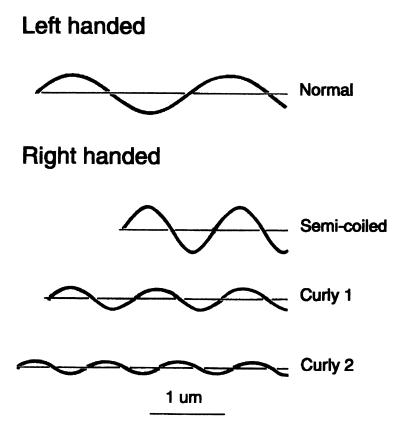
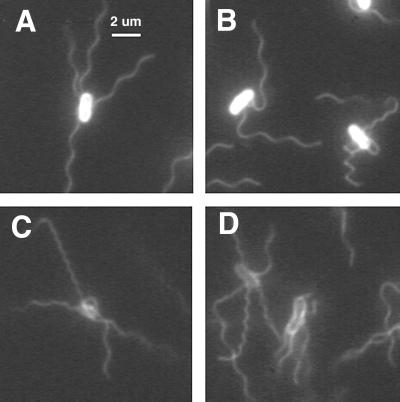
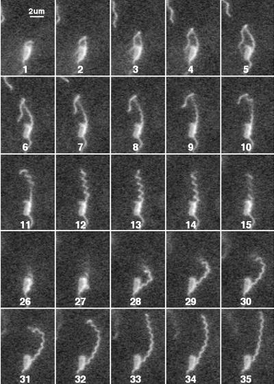
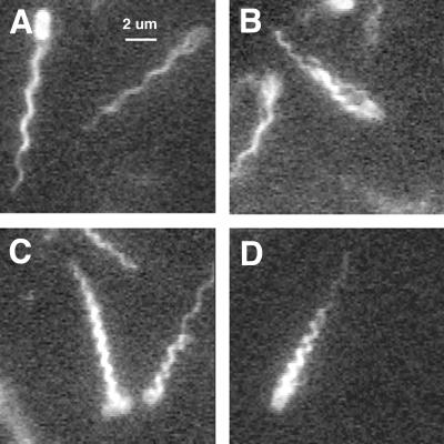
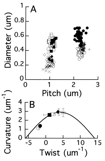
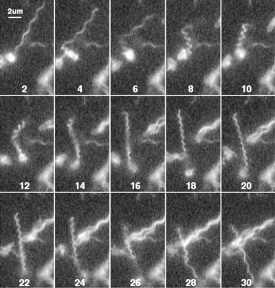
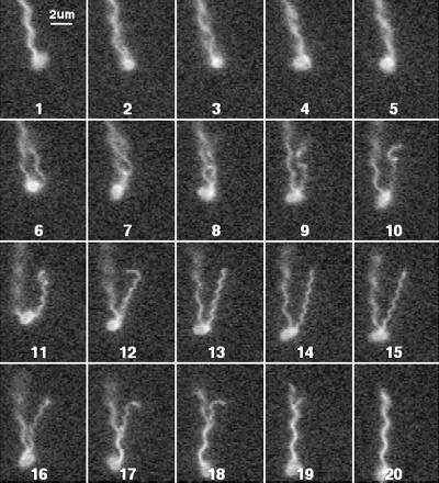
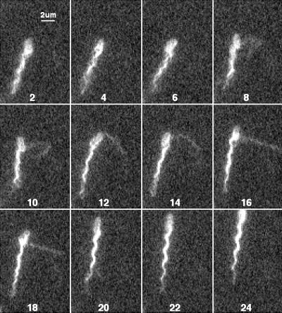
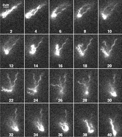
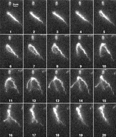
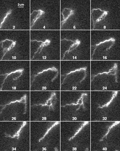
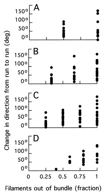
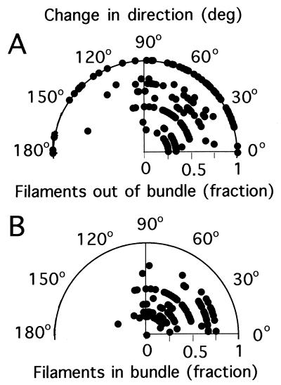
Bacteria swim by rotating flagellar filaments that are several micrometers long, but only about 20 nm in diameter. The filaments can exist in different polymorphic forms, having distinct values of curvature and twist. Rotation rates are on the order of 100 Hz. In the past, the motion of individual filaments has been visualized by dark-field or differential-interference-contrast microscopy, methods hampered by intense scattering from the cell body or shallow depth of field, respectively. We have found a simple procedure for fluorescently labeling cells and filaments that allows recording their motion in real time with an inexpensive video camera and an ordinary fluorescence microscope with mercury-arc or strobed laser illumination. We report our initial findings with cells of Escherichia coli. Tumbles (events that enable swimming cells to alter course) are remarkably varied. Not every filament on a cell needs to change its direction of rotation: different filaments can change directions at different times, and a tumble can result from the change in direction of only one. Polymorphic transformations tend to occur in the sequence normal, semicoiled, curly 1, with changes in the direction of movement of the cell body correlated with transformations to the semicoiled form.


Figures

FIG. 1
FIG. 1
Drawing of four different flagellar waveforms, each with a contour length of 4 μm. A filament of this length contains about 8,000 molecules of flagellin (12). The normal filament is left-handed, and the semicoiled, curly 1, and curly 2 filaments are right-handed. The normal and curly 1 filaments have the same overall length. Bar, 1 μm. Adapted from Calladine (7).
FIG. 2
FIG. 2
Immobilized cells of E. coli viewed for 1 s. (A and B) Labeled with Oregon Green 514 and illuminated by a mercury arc. (C and D) Labeled with Alexa Fluor 532 and illuminated by a strobed argon-ion laser (the technique used for all subsequent figures). The waveforms exhibited by individual filaments include normal (A); normal, semicoiled, and curly 1 (B); and normal, curly 1, and curly 2 (C and D).
FIG. 3
FIG. 3
Immobilized cell with a rotating filament undergoing polymorphic transformations. Successive fields are shown at 60 Hz (deinterlaced; total time span, 0.57 s). Fields 16 to 25 looked like field 26 and have been omitted.
FIG. 4
FIG. 4
Swimming cells with different kinds of flagellar bundles. Single fields are shown (deinterlaced). The waveforms of the flagellar bundles are normal (A), normal or curly 1 (both loose) (B), curly 1 (tight, but with one of the filaments on the cell at the right with a normal distal segment) (C), and semicoiled (with one filament with a normal distal segment) (D).
FIG. 5
FIG. 5
(A) Measurements of the diameter and pitch of stationary flagellar filaments (as in Fig. 2) and of bundles in swimming cells (as in Fig. 4). Filaments: ○, normal; +, normal de-energized with FCCP; □, semicoiled; ◊, curly 1; X, curly 2. Bundles: ●, normal; ■, semicoiled; ⧫, curly 1. (B) For each filament or bundle, curvature and twist were calculated from the diameter and pitch, and the mean values and SDs in these values were plotted. Symbols are the same as in panel A.
FIG. 6
FIG. 6
E. coli cell with one flagellar filament undergoing a polymorphic transformation. Every other field is shown.
FIG. 7
FIG. 7
E. coli with two flagellar filaments, one undergoing a polymorphic transformation. Successive fields are shown.
FIG. 8
FIG. 8
E. coli cell with several flagellar filaments, one undergoing a polymorphic transformation. Every other field is shown.
FIG. 9
FIG. 9
E. coli cell with several flagellar filaments, all but one undergoing polymorphic transformations. Every other field is shown.
FIG. 10
FIG. 10
E. coli cell with a bundle with a mixed waveform (curly 1 and normal) that becomes all normal, with some of the filaments leaving and then rejoining the bundle. Successive fields are shown.
FIG. 11
FIG. 11
E. coli cell with a normal bundle that transforms to a mixed waveform, where again filaments leave and rejoin the bundle. Every other field is shown.
FIG. 12
FIG. 12
Change in direction from run to run plotted as a function of the fraction of filaments out of the bundle. (A) Cells with two filaments. (B) Cells with three filaments. (C) Cells with four filaments. (D) Cells with five filaments.
FIG. 13
FIG. 13
Polar plots of the change in direction from run to run as a function of fraction of filaments out of the bundle (A) and fraction of filaments remaining in the bundle (B) (both plotted radially). Data are for cells with one to six filaments.

References

    1. Adler J, Templeton B. The effect of environmental conditions on the motility of Escherichia coli. J Gen Microbiol. 1967;46:175–184. - PubMed
    1. Armstrong J B, Adler J, Dahl M M. Nonchemotactic mutants of Escherichia coli. J Bacteriol. 1967;93:390–398. - PMC - PubMed
    1. Asakura S. Polymerization of flagellin and polymorphism of flagella. Adv Biophys. 1970;1:99–155. - PubMed
    1. Berg H C, Brown D A. Chemotaxis in Escherichia coli analysed by three-dimensional tracking. Nature. 1972;239:500–504. - PubMed
    1. Berg H C, Brown D A. Chemotaxis in Escherichia coli analyzed by three-dimensional tracking. Addendum. Antibiot Chemother. 1974;19:55–78. - PubMed

Publication types

MeSH terms

LinkOut - more resources