

MR digital subtraction angiography of cerebral arteriovenous malformations
- PMID: 10782782
- PMCID: PMC7976644
MR digital subtraction angiography of cerebral arteriovenous malformations
Abstract
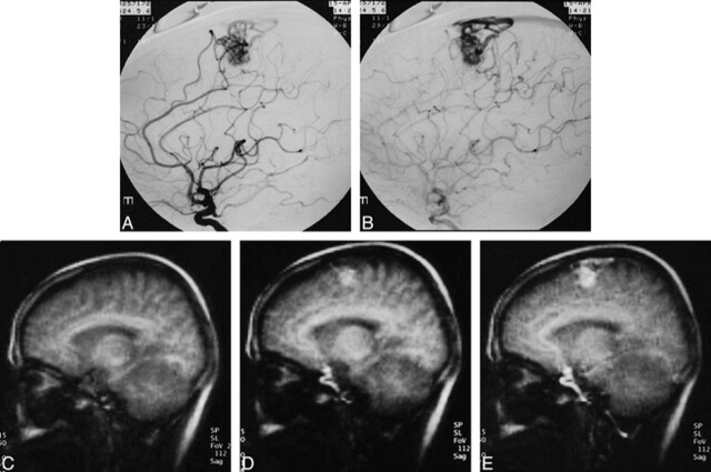
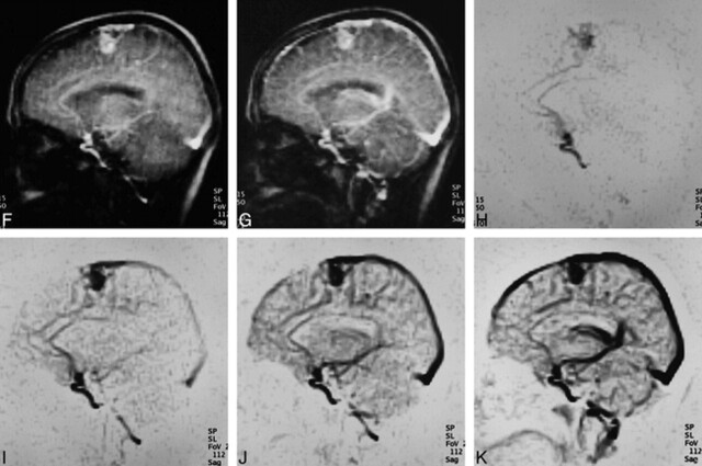
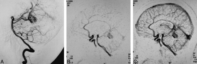
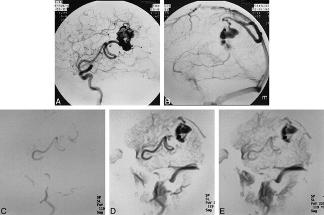
Background and purpose: Although phase-contrast MR angiography provides some information regarding hemodynamics of cerebral arteriovenous malformations (AVMs), most conventional MR angiographic techniques have not been helpful in this respect. We attempted to determine the value of MR digital subtraction angiography (DSA) in assessing AVM hemodynamics.
Methods: We developed an MR DSA technique by combining rapid thick-section T1-weighted imaging with a bolus injection of contrast material. The temporal resolution was 0.56 to 0.61 seconds per scan. MR DSA images obtained from 14 patients with AVMs were reviewed. Anatomic depiction of each component of the AVM was rated using a four-point grading scale (excellent = 3, good = 2, fair = 1, poor = 0) to compare conventional vs MR angiograms.
Results: We were able to obtain serial images in which passage of contrast material was evident within the AVM, although the sequence we used allowed images to be obtained in only one projection. The average score for feeders, nidi, and drainers was 1.6, 2.4, and 2.3, respectively, with an overall average of 2.1.
Conclusion: The spatial resolution of our technique may fall below the level needed for identification of small vascular components of an AVM. Additionally, the limited slab may restrict application of the technique to assessment of large or very small AVMs. MR DSA, however, can show the hemodynamics of AVMs and may serve as a supplement to conventional MR imaging in the diagnosis of cerebral AVMs.
Figures
References
-
- Huston J III, Rufenacht DA, Ehman RL, Wiebers DO. Intracranial aneurysms and vascular malformations: comparison of time-of-flight and phase-contrast MR angiography. Radiology 1991;181:721-730 - PubMed
-
- Marks MP, Pelc MJ, Ross MR, Enzmann DR. Determination of cerebral blood flow with a phase-contrast cine MR imaging technique: evaluation of normal subjects and patients with arteriovenous malformation. Radiology 1992;182:467-476 - PubMed
-
- Turski P, Korosec F. Phase contrast angiography. In: Anderson CM, Edelman R, Turski P, eds. Clinical Magnetic Resonance Angiography. New York: Raven; 1993:43-72
-
- Pant B, Sumida M, Arita K, Tominaga A, Ikawa F, Kurisu K. Usefulness of three dimensional phase contrast MR angiography on arteriovenous malformations. Neurosurg Rev 1997;20:171-176 - PubMed
-
- Marchal G, Michiels J, Bosmans H, Van Hecke P. Contrast-enhanced MRA of the brain. J Comput Assist Tomogr 1992;16:25-29 - PubMed
MeSH terms
LinkOut - more resources
Full Text Sources