

Case Reports
Carotid-cavernous fistulas: diagnosis with spiral CT angiography
Affiliations
- PMID: 10782783
- PMCID: PMC7976654
Item in Clipboard
Case Reports
Carotid-cavernous fistulas: diagnosis with spiral CT angiography
AJNR Am J Neuroradiol.
2000 Apr.
Abstract
Four cases in which the diagnosis of carotid-cavernous fistula was made by using CT angiography are illustrated. The diagnosis was confirmed by digital subtraction angiography in all four instances. To our knowledge, this is the first report of the CT angiographic appearance of carotid-cavernous fistulas.
Figures
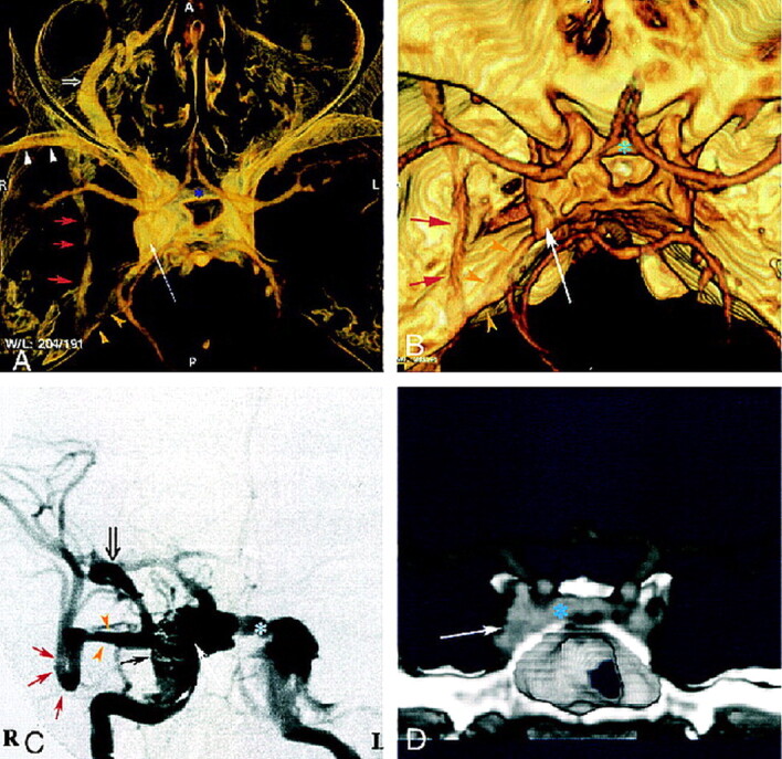
Case 1: 40-year-old man with direct, posttraumatic, right-sided CCF. A and B, Superior view of 3D CT angiogram obtained with the volume rendering technique shows an enlarged right cavernous sinus (long arrow ) with several draining veins: large right SOV (open arrow, A), anterior intercavernous sinus (asterisk ), inferior petrous sinus (yellow arrowheads), sphenoparietal sinus (white arrowheads, A), and paracavernous sinus (red arrows). C, DSA during embolization with selective right internal carotid artery injection shows right cavernous fistula with very high flow and multiple venous drainage channels, including large right SOV (open arrow ), anterior intercavernous sinus (asterisk ), sphenoparietal sinus (yellow arrowheads), and paracavernous sinus (red arrows). Closed black arrow indicates GDC coils; long white arrow, right cavernous sinus. D, Coronal view of 3D CT angiogram obtained with the volume rendering technique, using anterior cutting, clearly depicts intercavernous sinus (asterisk ). Arrow indicates right cavernous sinus.
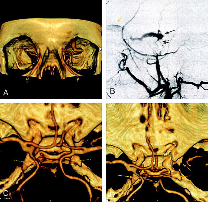
Case 2: 69-year-old woman with dural cavernous fistula. A, Frontal view of 3D CT angiogram obtained with the volume rendering technique shows an enlarged left SOV at the superior orbital fissure (single arrow ) and dilated angular veins (double arrows) bilaterally. B, DSA (selective left external carotid injection, arterial phase, lateral view) shows rapid opacification of a portion of the cavernous sinus (single black arrow ) supplied by middle meningeal artery branches (double black arrows) and draining anteriorly into an enlarged left SOV (yellow arrow ). C, 3D CT angiogram obtained with the volume rendering technique (superior view) shows bilateral enlarged cavernous sinuses (arrows). D, CT angiogram (superior view) better depicts the prominent left cavernous sinus (arrows) after cutting to exclude supracavernous internal carotid artery.
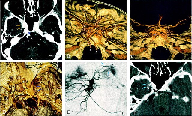
Case 3: 54-year-old woman with dural CCF and right exophthalmos, chemosis, and dilated episcleral vessels. A, CT angiogram (axial source image) shows an enlarged right cavernous sinus with irregular wall (yellow arrows) and large ipsilateral basilar plexus (asterisk ). White arrows indicate vertebral arteries. B and C, Superolateral (B ) and posterior (C ) views of 3D CT angiogram obtained with the volume rendering technique show inferior petrosal sinuses along the posterior surface of the petrous bone (yellow arrows). Note the good delineation of skull and vascular anatomy. Asterisk indicates basilar plexus; white arrows, vertebral arteries. D–F, Superolateral view of 3D CT angiogram obtained with the volume rendering technique (D ) shows enlarged cavernous sinus (yellow arrow ). Small vessels (blue arrows) might correspond to arteriovenous shunts, well depicted by lateral selective internal maxillary injection on DSA (E ). CT angiogram (axial source image) after superselective intraarterial embolization shows n-butyl cyanoacrylate in these arterial feeders (F ).
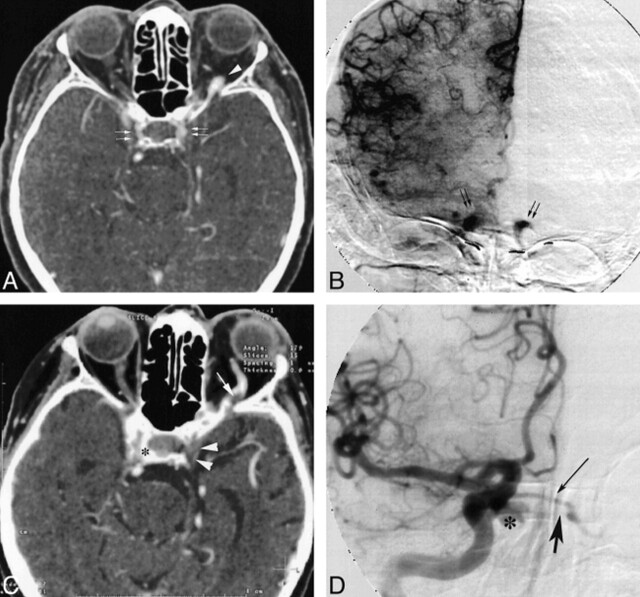
Case 4: 69-year-old woman with a dural fistula involving bilateral cavernous sinuses. A, CT angiogram (axial source image) shows enhancement of bilateral cavernous sinuses (double arrows) and enlarged left SOV (arrowhead ). B, DSA (right common carotid injection, late arterial phase, anteroposterior view) shows early opacification of bilateral enlarged cavernous sinuses (arrows). C, CT angiogram (axial source image), after partial embolization, shows partial thrombosis of left SOV (arrow ), without enhancement of left cavernous sinus (arrowheads), and persistent right enlarged cavernous sinus (asterisk ). D, DSA (right common carotid injection, arterial phase, anteroposterior view) confirms rapid opacification of right cavernous sinus (asterisk ) and intercavernous sinus (thin arrow ), without enhancement of left cavernous sinus (thick arrow ).
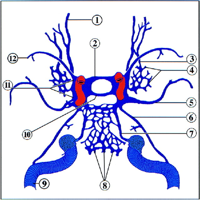
Schematic anatomic diagram of the venous vasculature of the skull base (superior view). 1, superior ophthalmic vein; 2, anterior intercavernous sinus; 3, inferior ophthalmic vein; 4, pterygoid plexus; 5, middle meningeal vein; 6, superior petrosal sinus; 7, inferior petrosal sinus; 8, basilar venous plexus; 9, transverse sinus; 10, posterior intercavernous sinus; 11, cavernous sinus; 12, sphenoparietal sinus
Similar articles
-
Dural carotico-cavernous fistula: pre and postembolization appearances of bone-subtracted CT angiography.Turk Neurosurg. 2013;23(2):249-51. doi: 10.5137/1019-5149.JTN.4301-11.1. Turk Neurosurg. 2013. PMID: 23546913
-
Classification and digital subtraction angiography evaluation of carotid cavernous fistulas.Chin Med J (Engl). 1999 Aug;112(8):735-8. Chin Med J (Engl). 1999. PMID: 11601284
-
Usefulness of C-arm cone-beam computed tomography in endovascular treatment of traumatic carotid cavernous fistulas: a technical case report.Neurosurgery. 2010 Aug;67(2):467-9; discussion 469-70. doi: 10.1227/01.NEU.0000372087.71176.FB. Neurosurgery. 2010. PMID: 20644434
-
[Spontaneous bilateral carotid-cavernous fistulas: about a case and review of the literature].Pan Afr Med J. 2017 Jun 6;27:91. doi: 10.11604/pamj.2017.27.91.8594. eCollection 2017. Pan Afr Med J. 2017. PMID: 28819512 Free PMC article. Review. French.
-
Spontaneous resolution of direct carotid-cavernous fistulas: case series and literature review.Interv Neuroradiol. 2019 Feb;25(1):71-89. doi: 10.1177/1591019918800220. Epub 2018 Sep 23. Interv Neuroradiol. 2019. PMID: 30244626 Free PMC article. Review.
Cited by
-
Clinically Directed Neuroimaging of Ophthalmoplegia.Clin Neuroradiol. 2018 Mar;28(1):3-16. doi: 10.1007/s00062-017-0646-0. Epub 2017 Nov 17. Clin Neuroradiol. 2018. PMID: 29149358 Review.
-
Traumatic carotid-cavernous fistula: excellent demonstration on 3D CT angiography.BMJ Case Rep. 2013 Oct 16;2013:bcr2013201707. doi: 10.1136/bcr-2013-201707. BMJ Case Rep. 2013. PMID: 24132453 Free PMC article. No abstract available.
-
Endovascular treatment of carotid cavernous sinus fistula: A systematic review.World J Radiol. 2013 Apr 28;5(4):143-55. doi: 10.4329/wjr.v5.i4.143. World J Radiol. 2013. PMID: 23671750 Free PMC article.
-
Trabeculae in the basilar venous plexus: anatomical and histological study with application to intravascular procedures.Anat Cell Biol. 2023 Dec 31;56(4):435-440. doi: 10.5115/acb.23.171. Epub 2023 Oct 17. Anat Cell Biol. 2023. PMID: 37845177 Free PMC article.
-
Feasibility of Noninvasive Diagnosis and Treatment Planning in a Case Series with Carotid-Cavernous Fistula using High-Resolution Time-Resolved MR-Angiography with Stochastic Trajectories (TWIST) and Extended Parallel Acquisition Technique (ePAT 6) at 3 T.Clin Neuroradiol. 2015 Sep;25(3):241-7. doi: 10.1007/s00062-014-0298-2. Epub 2014 Mar 6. Clin Neuroradiol. 2015. PMID: 24599323
References
-
- Leclerc X, Godefroy O, Pruvo JP, Leys D. Computed tomographic angiography for the evaluation of carotid artery stenosis. Stroke 1995;26:1577-1581 - PubMed
-
- Simonetti G, Bozzao A, Floris R, Silvestrini M. Non-invasive assessment of neck-vessel pathology. Eur Radiol 1998;8:691-697 - PubMed
-
- Leclerc X, Godefroy O, Lucas C, et al. Internal carotid arterial stenosis: CT angiography with volume rendering. Radiology 1999;210:673-682 - PubMed
-
- Schwartz RB, Tice HM, Hooten SM, Hsu L, Stieg PE. Evaluation of cerebral aneurysms with helical CT: correlation with conventional angiography and MR angiography. Radiology 1994;192:717-722 - PubMed
Publication types
MeSH terms
LinkOut - more resources
Full Text Sources
Medical