

MR imaging of patients with carotidynia
- PMID: 10782793
- PMCID: PMC7976649
MR imaging of patients with carotidynia
Abstract
Background and purpose: Carotidynia is an idiopathic neck pain syndrome associated with tenderness to palpation over the carotid bifurcation. Although well known in the otolaryngology and neurology literature, the validity of the entity has recently been questioned, in part because of the almost uniform absence of radiologic or pathologic findings. We report the MR findings in five patients with carotidynia.
Methods: During a period of 44 months, five patients with clinical signs and symptoms consistent with carotidynia were referred for imaging from the otolaryngology service. Each patient underwent MR imaging of the neck on a 1.5-T system. The studies included, as a minimum, pre- and postcontrast axial and postcontrast coronal T1-weighted images. Two patients also had axial T2-weighted imaging and another two patients underwent duplex sonography of the carotids.
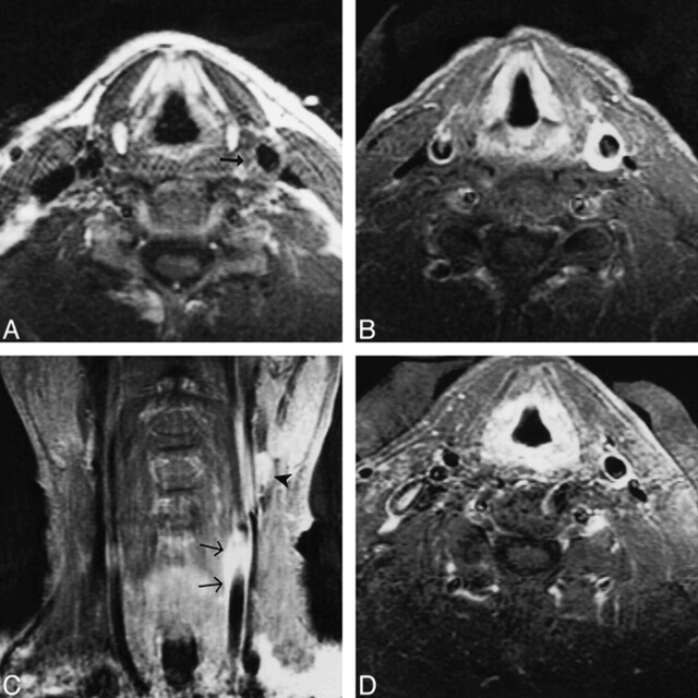
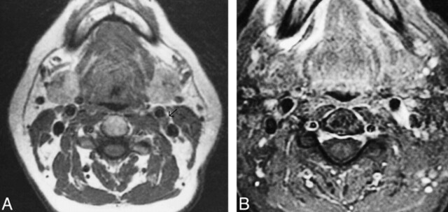
Results: All five patients had abnormal enhancing tissue surrounding the symptomatic carotid artery centered at the level of the distal common carotid and carotid bifurcation. This tissue had intermediate signal intensity on T1-weighted images and showed marked enhancement. In all patients, the remaining visualized portions of the carotid artery were normal. Normal flow voids were present throughout the vessel, and the caliber of the vessels was always within normal limits. There was no evidence of intramural hematoma, cervical lymphadenopathy, or atherosclerotic disease of the vessel. In one patient, repeat imaging after resolution of symptoms showed an absence of the previous abnormality.
Conclusion: The MR findings in these patients, along with the lack of any findings to suggest alternative diagnoses, support the existence of carotidynia as a distinct clinical entity.
Figures

References
-
- Fay T. Atypical neuralgia. Arch Neurol Psychiatry 1927;18:309-315
-
- Hilger JA. Carotid pain. Laryngoscope 1949;59:829-838 - PubMed
-
- Roseman DM. Carotidynia, a distinct syndrome. Arch Otolaryngol 1967;85:81-84 - PubMed
-
- Chiossone E, Quiroga ME. Carotidynia. J Laryngol Otol 1973;87:885-890 - PubMed
-
- Lovshin LL. Vascular neck pain: a common syndrome seldom recognized. Clev Clin Q 1960;27:5-13 - PubMed
MeSH terms
LinkOut - more resources
Full Text Sources
Medical