

Case Reports
Imaging of nonlaryngeal neuroendocrine carcinoma
Affiliations
- PMID: 10782795
- PMCID: PMC7976639
Item in Clipboard
Case Reports
Imaging of nonlaryngeal neuroendocrine carcinoma
AJNR Am J Neuroradiol.
2000 Apr.
Abstract
The imaging and pathologic features of three cases of nonlaryngeal neuroendocrine carcinoma of the head and neck are described. Neuroendocrine carcinomas represent malignant epithelial neuroendocrine neoplasms and are classified as three types: typical carcinoid (well differentiated), atypical carcinoid (moderately differentiated), and small cell neuroendocrine (poorly differentiated) carcinomas. The CT and MR imaging features of these tumors are nonspecific. Paranasal sinus neuroendocrine carcinomas showed expansion and destruction of the sinus, whereas metastatic neuroendocrine carcinomas to an intraparotid lymph node presented as a circumscribed parotid mass on CT scans.
Figures
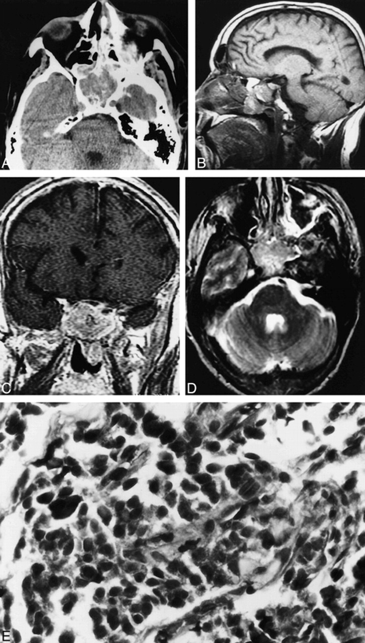
Case 1. A, Axial unenhanced CT scan shows a soft-tissue density mass in the sphenoid sinus. The mass is seen to expand the sinus. B, Sagittal T1-weighted MR image (600/16/1 [TR/TE/excitations]) shows the nasopharyngeal component of the sphenoid mass. Note that the floor of the sella appears intact. C, Coronal contrast-enhanced T1-weighted MR image (433/17/1) shows mild heterogeneous enhancement of the sphenoid sinus mass. D, Axial fast spin-echo T2-weighted MR image (3250/95/3) shows the sphenoid sinus mass to be heterogeneously hyperintense. E, Neuroendocrine carcinoma of the sphenoid sinus consisting of small pleomorphic cells exhibiting crowded hyperchromatic nuclei (hematoxylin and eosin stain; original magnification, ×40).
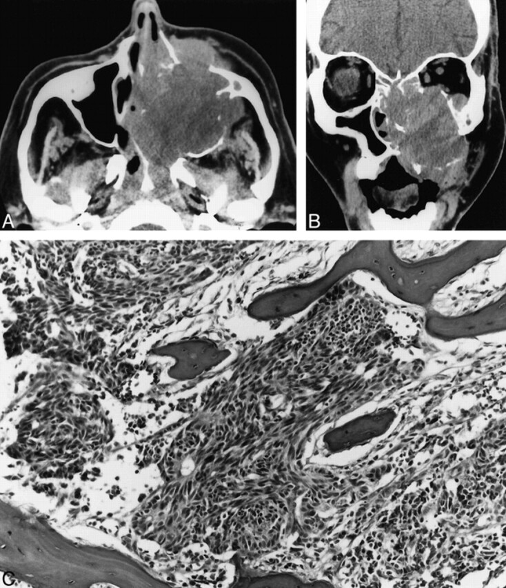
Case 2. A, Axial unenhanced CT scan shows a large expansile maxillary sinus mass with extension into the nasal cavity, the nasopharynx, the infratemporal fossa, and the subcutaneous tissues of the left maxillary region. B, Coronal unenhanced CT scan shows a large expansile mass in the left maxillary sinus that erodes the sinus walls and extends into the nasal cavity, left orbit, and subcutaneous soft tissues of the maxillary region. C, Poorly differentiated neuroendocrine carcinoma infiltrating maxillary bone. The tumor consists of small pleomorphic spindled cells with hyperchromatic nuclei. Focal rosette formation is noted (hematoxylin and eosin stain; original magnification, ×10).
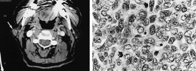
Case 3. A, Axial contrast-enhanced CT scan shows a mildly enhancing circumscribed mass (arrow) in the lateral aspect of the superficial lobe of the right parotid gland. B, Metastatic poorly differentiated neuroendocrine carcinoma to an intraparotid lymph node. The tumor cells display pleomorphic vesicular nuclei with finely dispersed chromatin, scant cytoplasm, and a high mitotic rate (hematoxylin and eosin stain; original magnification, ×40).
References
-
- Batsakis JG, El-Naggar AK, Luna MA. Neuroendocrine tumors of the larynx. Ann Otol Rhinol Laryngol 1992;101:710-714 - PubMed
-
- Shanmugaratnam K, Sobin LH, Barnes L, et al. Histological Typing of Tumors of the Upper Respiratory Tract and Ear, World Health Organization (International Histological Classification of Tumors). 2nd ed. New York: Springer-Verlag; 1991
-
- Wenig BM, Hyams VJ, Heffner DK. Moderately differentiated neuroendocrine carcinoma of the larynx. Cancer 1998;62:2658-2676 - PubMed
-
- Erlandson RA, Nesland JM. Tumors of the endocrine/neuroendocrine system. Ultrastruct Pathol 1994;18:149-170 - PubMed
-
- El-Nagger AK, Batsakis JG. Carcinoid tumor of the larynx: a critical review of the literature. ORL J Otorhinolaryngol Relat Spec 1991;53:188-193 - PubMed
Publication types
MeSH terms
LinkOut - more resources
Full Text Sources
Medical