. 2000 Apr 15;14(8):913-26.

Mcm10 and the MCM2-7 complex interact to initiate DNA synthesis and to release replication factors from origins

Affiliations

Mcm10 and the MCM2-7 complex interact to initiate DNA synthesis and to release replication factors from origins

L Homesley et al. Genes Dev. .

Abstract

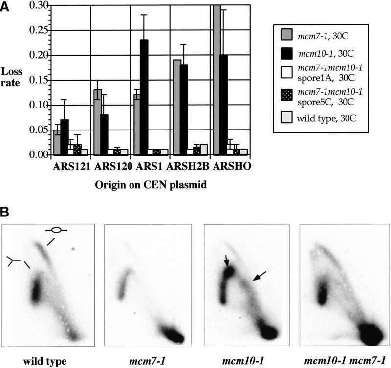
MCM2-7, a complex of six subunits, is an essential component of the prereplication chromatin that is assembled at Saccharomyces cerevisiae replication origins during G(1) phase. It is also believed to be the processive helicase at growing forks. To elucidate the action of MCM2-7 during the transition from initiation to elongation replication, we have focused our studies on Mcm10, a replication initiation protein that physically interacts with members of the MCM2-7 complex. We show that Mcm10 is a chromatin-associated protein that mediates the association of the MCM2-7 complex with replication origins. Furthermore, diminished interaction between Mcm10 and Mcm7, a subunit of the MCM2-7 complex, by a mutation in either Mcm10 or Mcm7 inhibits replication initiation. Surprisingly, a double mutant containing both the mcm10-1 and mcm7-1 (cdc47-1) alleles restores interaction between Mcm10 and Mcm7 and corrects all of the defects exhibited by each of the single mutants, including the stalling of replication forks at replication origins typically seen in mcm10-1 cells. This mutual compensation of defects between two independently isolated mutations is allele specific. These results suggest that Mcm10, like Mcm7, is a critical component of the prereplication chromatin and that interaction between Mcm10 and Mcm7 is required for proper replication initiation and prompt release of origin-bound factors.


Figures

Figure 1
Figure 1
Two-dimensional DNA gel analysis of replicative intermediates in the ORI1 region in the mcm10-1 mutant. (A) ORI1 in a wild-type strain. (B) ORI1 in the mcm10-1 mutant strain. The cartoon is an interpretation of the result showing that the intense spots are due to accumulations of specific species of replicative intermediates. (C) The ori1 alleles in the mcm10-1 mutants: (a) ori1-a; (b) ori1-b2b3; (c) ori1-b1; (d) ori1-b3. Pause signals are indicated by open triangles. All ori1 alleles were constructed in isogenic mcm10-1 background.
Figure 2
Figure 2
Mcm10 is totally and constitutively bound to chromatin. (A) Western blot analysis of proteins in whole cell extracts (W), soluble fractions (S), and chromatin pellets (P) from cells arrested at either G1, S, or M phases. (Top) Mcm10; (middle) actin; (bottom) histone H2B. (B) Association of Mcm10 and Mcm2 with chromatin during the time course of one cell cycle. Cells were harvested at 10-min intervals after being released from α factor synchronization. The chromatin pellet of each sample was analyzed in SDS-PAGE and immunoblots were probed with anti-Mcm10 (top) and anti-Mcm2 (bottom) antibodies. (C) Association of Mcm10 with chromatin is DNA dependent. Whole cell extracts were treated at 37°C for 10 min with DNase I (500 U/ml; lanes 1,2), RNase A (50 mg/ml; lanes 3,4), NP-40 (2%; lanes 5,6), or lysis buffer (lanes 7,8), then fractionated into soluble (S) and pellet (P) fractions by centrifugation. Proteins in the pellets (lanes 1,3,5,7) and soluble fractions (lanes 2,4,6,8) were separated by SDS-PAGE. Immunoblots were probed with anti-Mcm10 (top), and anti-histone H2B (bottom) antibodies. Chromatin fractionation was performed essentially as described (Yan et al. 1991; Liang et al. 1995; Donovan et al. 1997) except that a low speed (4000 rpm) centrifugation was performed after the cells were lysed to remove unbroken cells. The yeast strain used in A, B, and C is W303bar1. (D) Mcm10 is localized to ORI1 by in vivo cross-linking. PCR products from crude extracts of Mcm10 (lane 1) and Mcm10–Myc strain (lane 6) as well as immunoprecipitates from the Mcm10 (lanes 25) and Mcm10–Myc strain (lanes 710) were analyzed on PAG. Samples treated with formaldehyde (lanes 4,5,9,10), or anti-Myc antibodies (lanes 2,3,7,8) are indicated. R2.5 is a 240-bp DNA fragment located 2.5 kb from ORI1.
Figure 3
Figure 3
Mcm10 is required for the binding of the MCM2–7 complex to chromatin. (A) Chromatin binding of Mcm10, Orc3, Mcm2, and histone H2B during G1 phase in MW23 cells (a ura3-53 can1-11 mcm10-43) (Solomon et al. 1992). Cells were treated with α factor (10 μg/ml) for 3 hr at 30°C and again with the same amount before being shifted to 37°C for 0–5 hr (lanes 15). Pellet (P) and soluble fractions (S) were prepared as described in Fig. 2, and analyzed in SDS-PAGE. Western blots were probed with antibodies specific for Mcm10, Orc3, Mcm2, or histone H2B as indicated. (B) Chromatin binding of Orc3, Mcm10, Mcm2, and histone H2B during G1 phase in JRY4490 cells (a can1-1 his3-11 leu2-3, -112 lys2Δ trp1-1 ura3-1 orc2-1) (Foss et al. 1993). Cells were treated with α factor (10 μg/ml) for 3 hr at 25°C and again with the same amount before being shifted to 37°C for 0–5 hr (lanes 15). Pellet and soluble fractions were analyzed in SDS-PAGE. Western blots were probed with anti-Mcm10, anti-Orc3, anti-Mcm2, or anti-histone H2B as indicated. The anti-Orc3 antibody was a gift from Bruce Stillman (Cold Spring Harbor Laboratory) and the anti-H2B antibody was a gift from Mike Grunstein (UCLA). (C) Mcm10, Mcm2, and Orc2 are associated with chromatin in the presence of α factor after 5 hr of incubation at 37°C in the W303bar1 strain.
Figure 4
Figure 4
Tetrad analyses of diploids from a cross between R125A22-2C (mcm10-1) and DBY2029 (mcm7-1). (A) Segregation patterns of phenotypes indicate that the double mutant is wild type, although both parental strains are temperature sensitive (ts). (TT) Tetratype; (PD) parental ditype; (NPD) nonparental ditype. Both parental strains are ts. (B) Growth phenotypes of spores from typical tetrads from a cross between mcm10-1 and mcm7-1. Viable spores of a TT tetrad were further analyzed by crossing with a wild-type strain. (C) Growth phenotypes of yeast strains LHY1A (mcm10-1 mcm7-1), LHY4B (mcm10-1 mcm5-461), and LHYTM18C (mcm10-1 mcm7-1 mcm5-461) after 3 days on YEPD plates at 30°C and 37°C. (D) Growth rates of the single, double, and triple mutants of mcm10-1, mcm5-461, and mcm7-1. Strains were grown overnight in YPD at 30°C, then diluted to OD600 = 0.1. Diluted cultures were kept at 30°C for 30 min then shifted to 37°C for 10 hr. Doubling times were calculated from growth curves. The mcm7-1 strain did not grow after shift to 37°C.
Figure 5
Figure 5
Replication defects of the mcm10-1 and mcm7-1 single mutants are corrected in mcm10-1 mcm7-1. (A) Stability of plasmids in single and double mutants of mcm10-1 and mcm7-1. Plasmid loss rates were determined for the single mutants mcm7-1 and mcm10-1, as well as for two double mutants (LHY1A and LHY5C) and a wild-type strain (8534-8c) at 30°C. Five different minichromosomes each containing a different ARS were used in this assay. The corresponding ARS of each minichromosome (e.g., ARS121, ARS120) is indicated on the x-axis. The y-axis shows the rates of plasmid loss per cell per generation. (B) The double mutant relieves the pause phenotype of mcm10-1. Two-dimensional DNA gel analysis of mcm10-1 mcm7-1. Genomic DNA was isolated from the wild-type (8534-8c), mcm10-1, mcm7-1, and mcm10-1 mcm7-1 strains. Genomic DNA was digested with NcoI, separated on two-dimensional gel, blotted, and probed with 32P-labeled ORI1 DNA. Bubble and Y arcs are indicated by cartoons on the blot showing the DNA from the wild-type strain. Pause signals that result from accumulation of specific species of replication intermediates are indicated by arrows.
Figure 6
Figure 6
Interactions between wild-type and mutant Mcm7 and Mcm10 proteins. (A) Model to explain the mutual suppression of two mutant proteins by compensatory mutations that restore physical interactions. (Wild-type proteins) X and Y; (mutant proteins) x and y. (B) Physical interactions between wild-type and mutant proteins of Mcm10 and Mcm7 measured by β-galactosidase activity using the two-hybrid system. Mcm7 is fused to the Gal4 activation domain and Mcm10 is fused to the Gal4-binding domain. (C) Same as B with bait and prey in reverse orientations. The Mcm10-43 mutant protein is also included in the analysis. (D) Tetrad analyses of diploids from a cross between DBY2029 (mcm7-1) and mcm10-43. mcm7-1 was crossed with mcm10-43 in two different backgrounds (BTY106 and BTY103), and data were combined. The inferred genotypes indicate that the double mutant is temperature sensitive (ts). (TT) Tetratype; (PD) parental ditype; (NPD) nonparental ditype; (ts) temperature sensitive; (wt) wild-type phenotype. Both parental strains are ts. (E) Evolutionary conservation of the Mcm10 protein. The mcm10-1 and mcm10-43 mutations are indicated above the aligned sequences of putative homologs of Mcm10: (Cdc23) S. pombe; (Con4-2907) C. albicans; (WPY47D3) C. elegans. Both mutations occur in regions conserved between S. cerevisiae and S. pombe. Black boxes indicate consensus. Gray boxes indicate similarity to consensus. Consensus zinc finger motif is shown at the bottom.
Figure 6
Figure 6
Interactions between wild-type and mutant Mcm7 and Mcm10 proteins. (A) Model to explain the mutual suppression of two mutant proteins by compensatory mutations that restore physical interactions. (Wild-type proteins) X and Y; (mutant proteins) x and y. (B) Physical interactions between wild-type and mutant proteins of Mcm10 and Mcm7 measured by β-galactosidase activity using the two-hybrid system. Mcm7 is fused to the Gal4 activation domain and Mcm10 is fused to the Gal4-binding domain. (C) Same as B with bait and prey in reverse orientations. The Mcm10-43 mutant protein is also included in the analysis. (D) Tetrad analyses of diploids from a cross between DBY2029 (mcm7-1) and mcm10-43. mcm7-1 was crossed with mcm10-43 in two different backgrounds (BTY106 and BTY103), and data were combined. The inferred genotypes indicate that the double mutant is temperature sensitive (ts). (TT) Tetratype; (PD) parental ditype; (NPD) nonparental ditype; (ts) temperature sensitive; (wt) wild-type phenotype. Both parental strains are ts. (E) Evolutionary conservation of the Mcm10 protein. The mcm10-1 and mcm10-43 mutations are indicated above the aligned sequences of putative homologs of Mcm10: (Cdc23) S. pombe; (Con4-2907) C. albicans; (WPY47D3) C. elegans. Both mutations occur in regions conserved between S. cerevisiae and S. pombe. Black boxes indicate consensus. Gray boxes indicate similarity to consensus. Consensus zinc finger motif is shown at the bottom.
Figure 7
Figure 7
(A) Genetic interactions between mcm10-1, cdc45-1, mcm5-461 (cdc46-1), and mcm7-1 (cdc47-1). mcm5-461 and mcm7-1 are synthetic lethal mutations that were originally isolated as suppressors of the cold-sensitive cdc45-1 mutation (Hennessy et al. 1991; Moir et al. 1982). Arrow indicates suppression, double-headed arrow indicates mutual suppression, bar indicates synthetic lethality. (B) Genetic spore segregation patterns of tetrads derived from a cross between mcm10-1 and cdc45-1. Inferred spore types are shown in parentheses. (C) A model to explain the observations that allele-specific suppressors of mcm10-1 in the MCM2–7 gene family are also suppressors of cdc45-1. We hypothesize that the site on Mcm7 that contacts Mcm10 is also used to contact Cdc45, although these contacts may not occur at the same time. (a) The association of the MCM2–7 complex with a replication origin is mediated through Mcm10 by interactions with Mcm7 and possibly other subunits of the complex during G1 phase. Cdc45 is recruited to the pre-RC just before the G1 phase to S-phase transition (Zou and Stillman 1998). (b) In this model, displacement of MCM2–7 from Mcm10 by Cdc45 is a critical step in the activation of replication initiation. Once initiation has occurred, Cdc45 and MCM2–7 may comigrate from the replication origin in concert with the elongation machinery (Aparicio et al. 1997). The large gray oval shape in a and b represents, respectively, the pre-RC and post-RC in transition.

Similar articles

Cited by

References

    1. Alani E, Cao L, Kleckner N. A method for gene disruption that allows repeated use of URA3 selection in the construction of multiply disrupted yeast strains. Genetics. 1987;116:541–545. - PMC - PubMed
    1. Aparicio OM, Weinstein DM, Bell SP. Components and dynamics of DNA replication complexes in S. cerevisiae: Redistribution of MCM proteins and Cdc45p during S phase. Cell. 1997;91:59–69. - PubMed
    1. Ausubel FM, Brent T, Kingston RE, Moore DD, Seidman JG, Smith JA, Struhl K. Current protocols in molecular biology. Harvard, MA: John Wiley & Sons; 1998.
    1. Aves S, Tongue N, Foster AJ. The essential Schizosaccharomyces pombe cdc23 DNA replication gene shares structural and functional homology with the Saccharomyces cerevisiae DNA43 (MCM10) gene. Curr Genet. 1998;34:164–171. - PubMed
    1. Bell S, Stillman B. ATP-dependent recognition of eukaryotic origins of DNA replication by a multiprotein complex. Nature. 1992;357:128–134. - PubMed

Publication types

MeSH terms