. 2000 Apr 15;14(8):927-39.

BASC, a super complex of BRCA1-associated proteins involved in the recognition and repair of aberrant DNA structures

Affiliations

BASC, a super complex of BRCA1-associated proteins involved in the recognition and repair of aberrant DNA structures

Y Wang et al. Genes Dev. .

Abstract

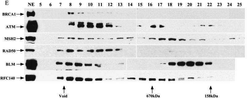
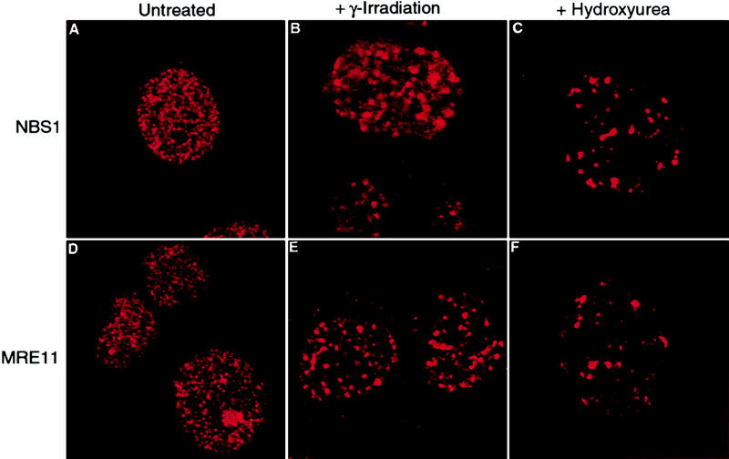
We report the identities of the members of a group of proteins that associate with BRCA1 to form a large complex that we have named BASC (BRCA1-associated genome surveillance complex). This complex includes tumor suppressors and DNA damage repair proteins MSH2, MSH6, MLH1, ATM, BLM, and the RAD50-MRE11-NBS1 protein complex. In addition, DNA replication factor C (RFC), a protein complex that facilitates the loading of PCNA onto DNA, is also part of BASC. We find that BRCA1, the BLM helicase, and the RAD50-MRE11-NBS1 complex colocalize to large nuclear foci that contain PCNA when cells are treated with agents that interfere with DNA synthesis. The association of BRCA1 with MSH2 and MSH6, which are required for transcription-coupled repair, provides a possible explanation for the role of BRCA1 in this pathway. Strikingly, all members of this complex have roles in recognition of abnormal DNA structures or damaged DNA, suggesting that BASC may serve as a sensor for DNA damage. Several of these proteins also have roles in DNA replication-associated repair. Collectively, these results suggest that BRCA1 may function as a coordinator of multiple activities required for maintenance of genomic integrity during the process of DNA replication and point to a central role for BRCA1 in DNA repair.


Figures

Figure 1
Figure 1
Immunoprecipitation of BRCA1-associated proteins and identification by mass spectrometry. (A) Immunoprecipitates of BRCA1-associated proteins using antibodies Ab80 and Ab81 were resolved on a 4%–20% gradient SDS–polyacrylamide gel and stained with Coomassie blue. Labeled protein bands were identified by mass spectrometry. (B) A representative MS/MS spectrum that identifies the 160-kD band from Ab80 immunoprecipitate as the RecQ DNA helicase BLM.
Figure 1
Figure 1
Immunoprecipitation of BRCA1-associated proteins and identification by mass spectrometry. (A) Immunoprecipitates of BRCA1-associated proteins using antibodies Ab80 and Ab81 were resolved on a 4%–20% gradient SDS–polyacrylamide gel and stained with Coomassie blue. Labeled protein bands were identified by mass spectrometry. (B) A representative MS/MS spectrum that identifies the 160-kD band from Ab80 immunoprecipitate as the RecQ DNA helicase BLM.
Figure 2
Figure 2
BRCA1-associated proteins that form a BASC. (A–D) Components of the BASC coimmunoprecipitate. Immunoprecipitations were done with HeLa nuclear extracts (NE) (A–C) and 293T whole-cell extracts (TCL) (D). Antibodies and immunoprecipitation/Western conditions are described in the Materials and Methods section. (E) BRCA1 resides in a large complex of >2 MD. Components of the BASC complex cofractionate on DEAE and Superose 6 columns. HeLa nuclear extracts were fractionated and step eluted (0.2–0.4 m KCl) on a DEAE column. The majority of BRCA1 eluted in the 0.3 m fraction. The 0.3 m fraction was fractionated further on a Superose 6 gel filtration column. BRCA1 was detectable only in the void volume (>2 MD). Other components of BASC were detected as multiple peaks; all contain one peak in the void volume that co-elutes with BRCA1. Other peaks of smaller sizes exist independent of BRCA1. The majority of the RAD50 complex that is independent of BRCA1 elutes in the 0.2 m KCl on the DEAE column.
Figure 2
Figure 2
BRCA1-associated proteins that form a BASC. (A–D) Components of the BASC coimmunoprecipitate. Immunoprecipitations were done with HeLa nuclear extracts (NE) (A–C) and 293T whole-cell extracts (TCL) (D). Antibodies and immunoprecipitation/Western conditions are described in the Materials and Methods section. (E) BRCA1 resides in a large complex of >2 MD. Components of the BASC complex cofractionate on DEAE and Superose 6 columns. HeLa nuclear extracts were fractionated and step eluted (0.2–0.4 m KCl) on a DEAE column. The majority of BRCA1 eluted in the 0.3 m fraction. The 0.3 m fraction was fractionated further on a Superose 6 gel filtration column. BRCA1 was detectable only in the void volume (>2 MD). Other components of BASC were detected as multiple peaks; all contain one peak in the void volume that co-elutes with BRCA1. Other peaks of smaller sizes exist independent of BRCA1. The majority of the RAD50 complex that is independent of BRCA1 elutes in the 0.2 m KCl on the DEAE column.
Figure 3
Figure 3
Colocalization of BRCA1 with BLM before and after exposure to genotoxic agents. (A–F) Asynchronous, logarithmically growing MCF7 cells were fixed with methanol/acetone and stained with antibodies to BRCA1 (Ab-1, Calbiochem) and BLM followed by the appropriate FITC and Cy3-conjugated secondary antibodies. (G–L) Cells were treated with 1 mm HU for 6–8 hr followed by fixation and staining. (M–O) Cells were exposed to 12 Gy of γ-irradiation and incubated for 8 hr prior to fixation and staining. Confocal images were captured at 1260× magnification.
Figure 4
Figure 4
Cell cycle dependent colocalization of BRCA1 with the MRE11–RAD50–NBS1 complex after ionizing radiation. (A–C,M–O) Asynchronous MCF7 cells were fixed and stained with (A–C) anti-RAD50 and anti-BRCA1 or (M–O) anti-MRE11 and anti-BRCA1 antibodies. Asynchronous (D–F,P–R), G0/G1 arrested (G–I,S–U), or S-phase synchronized (J–L,V–X) MCF7 cells were exposed to 12 Gy of γ-irradiation then incubated for 5 hr prior to fixation and immunostaining. Appropriate FITC or Cy3-conjugated secondary antibodies were used for indirect visualization of the epitopes. Confocal images were captured on a Bio-Rad confocal microscope at 1260× magnification.
Figure 5
Figure 5
Relocalization of the RAD50–MRE11–NBS1 complex following exposure to ionizing radiation or HU is independent of BRCA1. Exponentially growing HCC1937 cells were either left untreated (A,D), exposed to 12 Gy of ionizing radiation followed by an 8-hr incubation (B,E), or treated with 1 mm HU for 8 hr (C,F). Cells were fixed and stained with antibodies to NBS1 (Novus) or MRE11 (Novus) as indicated. (A,D) Cells without foci; (B,C,E,F) cells that contain foci. Confocal images were captured at 1260× magnification. The percentage of cells with foci in each condition was determined by scoring cells as positive if they contained >10 foci. In each experiment, >200 cells were counted.
Figure 6
Figure 6
Colocalization of RAD50, MRE11, BRCA1, and BLM following treatment of cells with HU. Asynchronous MCF7 cells were treated with 1 mm HU for 4–8 hr. Cells were fixed and stained with the following antibodies: (A–C) anti-RAD50 (13B3, GeneTex) and anti-BRCA1 (Ab-2, Neomarkers); (D–F) anti-MRE11 (12D, GeneTex) and anti-BRCA1 (Ab-2 NeoMarkers); (G–I) anti-MRE11 (12D7 GeneTex); and affinity-purified polyclonal anti-BLM; (J–L) anti-RAD50 (13B3, GeneTex) and affinity-purified polyclonal anti-BLM. Confocal images were captured at 1260× magnification. It should be noted that the majority of cells after HU treatment did not appear significantly different from the untreated cells. However, ∼10% showed redistribution into nuclear foci as represented in these images.
Figure 7
Figure 7
Colocalization of BLM, BRCA1, and RAD50 with PCNA following treatment with HU. Cells were left either untreated or exposed to 1 mm HU for 7 hr. Then, cells were fixed by methanol/acetone treatment and stained by indirect immunofluorescence with the following primary antibodies: (A–F) anti-PCNA (PC10, Santa Cruz) and anti-BRCA1 (Ab-2, Neomarkers); (G–L) anti-PCNA (PC10, Santa Cruz) and anti-BLM; (M–R) anti-PCNA (FL261, Santa Cruz) and anti-RAD50 (GeneTex). Appropriate Cy3- or FITC-conjugated secondary antibodies were used and confocal images were captured at 1260× magnification. It should be noted that the majority of cells after HU treatment did not appear significantly different from the untreated cells. However, ∼10% showed redistribution into nuclear foci as represented in these images.

References

    1. Abbott DW, Thompson ME, Robinson-Benion C, Tomlinson G, Jensen RA, Holt JT. BRCA1 expression restores radiation resistance in BRCA1-defective cancer cells through enhancement of transcription-coupled DNA repair. J Biol Chem. 1999;274:18808–18812. - PubMed
    1. Alani E, Lee S, Kane MF, Griffith J, Kolodner RD. Saccharomyces cerevisiae MSH2, a mispaired base recognition protein, also recognizes Holliday junctions in DNA. J Mol Biol. 1997;265:289–301. - PubMed
    1. Allen JB, Zhou Z, Siede W, Friedberg EC, Elledge SJ. The SAD1/RAD53 protein kinase controls multiple checkpoints and DNA damage- induced transcription in yeast. Genes & Dev. 1994;8:2401–2415. - PubMed
    1. Banin S, Moyal L, Shieh S, Taya Y, Anderson CW, Chessa L, Smorodinsky NI, Prives C, Reiss Y, Shiloh Y, Ziv Y. Enhanced phosphorylation of p53 by ATM in response to DNA damage. Science. 1998;281:1674–1677. - PubMed
    1. Bennett RJ, Keck JL, Wang JC. Binding specificity determines polarity of DNA unwinding by the Sgs1 protein of S. cerevisiae. J Mol Biol. 1999;289:235–248. - PubMed

Publication types

MeSH terms