. 2000 Apr 15;14(8):994-1004.

Induction of medulloblastomas in p53-null mutant mice by somatic inactivation of Rb in the external granular layer cells of the cerebellum

Affiliations

Induction of medulloblastomas in p53-null mutant mice by somatic inactivation of Rb in the external granular layer cells of the cerebellum

S Marino et al. Genes Dev. .

Abstract

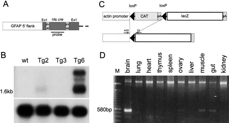
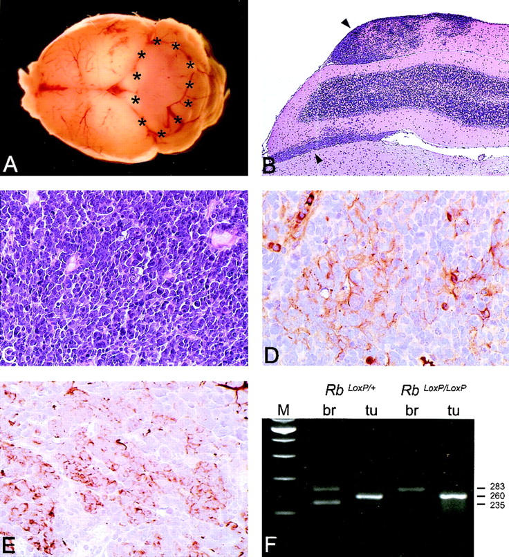
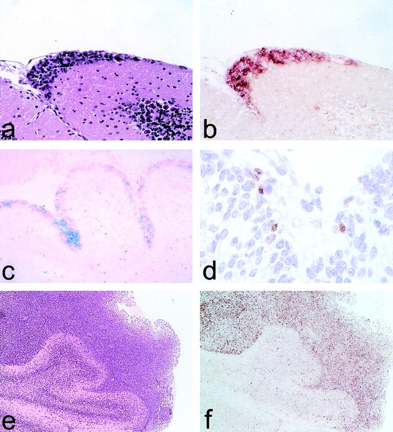
Medulloblastomas are among the most common malignancies in childhood, and they are associated with substantial mortality and morbidity. The molecular pathogenesis as well as the ontogeny of these neoplasms is still poorly understood. We have generated a mouse model for medulloblastoma by Cre-LoxP-mediated inactivation of Rb and p53 tumor suppressor genes in the cerebellar external granular layer (EGL) cells. GFAP-Cre-mediated recombination was found both in astrocytes and in immature precursor cells of the EGL in the developing cerebellum. GFAP-Cre;Rb(LoxP/LoxP);p53(-/- or LoxP/LoxP) mice developed highly aggressive embryonal tumors of the cerebellum with typical features of medulloblastoma. These tumors were identified as early as 7 weeks of age on the outer surface of the molecular layer, corresponding to the location of the EGL cells during development. Our results demonstrate that loss of function of RB is essential for medulloblastoma development in the mouse and strongly support the hypothesis that medulloblastomas arise from multipotent precursor cells located in the EGL.


Figures

Figure 1
Figure 1
Structure of GFAP–Cre transgene and characterization of the transgenic mice. (A) Partial structure of the GFAP–Cre transgene. (B) Northern blot analysis of Cre expression on poly(A)+ RNA from three independent transgenic lines. A 1.6-kb mRNA corresponding to the predicted transcript from the GFAP–Cre transgene was detected in Tg2 and Tg6 mice. Control hybridization with hGAPDH probe shows an equal loading of RNA in each lane. (C) Genomic structure of the transgene in the ACZL mice. Cre-mediated recombination leads to juxtaposition of the lacZ gene to the chicken β-actin promoter. (D) PCR analysis of recombination on GFAP–Cre;ACZL double transgenic mice. A 580-bp product for the recombined allele is present in brain, muscle (likely originating from peripheral myelinated nerve fibers), and intestine (enteric glia). The upper band represents the nonrecombined allele. The location of the primers AG2 and Z3 is illustrated in C.
Figure 2
Figure 2
In situ analysis of recombination in GFAP–Cre;ACZL double transgenic mice. (Upper row) Astrocytes double positive for LacZ staining (blue reaction product) and GFAP (brown stain) located in the brain stem (A) and in the cerebral cortex (B); double positive ependymal cells lining the ventricular wall (C) and neurons positive for LacZ staining and negative for GFAP in the CA4 portion of the neuronal ribbon of the hippocampus (D); double labeling of Cre mRNA (black) and GFAP protein (brown) in fibrillary white matter astrocytes. (E). (Lower row) Brain specimen 24 hr after cryolesioning. Macroscopical appearance of the lesioned brain (F) and histological analysis of the lesioned area on frontal sections stained with H&E (G) are shown. (H) Up-regulation of Cre expression in reactive astrocytes located in the necrotic area and surrounding the lesion as demonstrated by Cre mRNA in situ hybridization. In contrast, there is only little basal Cre expression in the cerebral cortex of an unlesioned animal (I).
Figure 3
Figure 3
Analysis of Rb and p53 recombination in different areas of the adult mouse brain. (A) Structure of the Rb floxed allele: LoxP sites were inserted into the introns surrounding exon 19. (B) A 260-bp PCR product representing the recombined Rb allele (primers Rb212 and Rb18) is present in a subset of cells in all analyzed areas of the brain. The 283-bp product represents the floxed allele (primers Rb19E and Rb18). The location of the primers used for the analysis (Rb212, Rb19E, and Rb18) is illustrated in A. Structure of the p53 floxed allele (C) and PCR analysis of recombination in different areas of the brain (D,E) are shown. The 584-bp band represents the floxed p53 allele (primers 10F and 10R), and the 612-bp band represents the recombined p53 allele (primers 1F and 10R). (M) Molecular marker; (T) tail; (OB) olfactory bulb; (Cx) cortex; (Hc) hyppocampus; (Ce) cerebellum; (Bs) brain stem.
Figure 3
Figure 3
Analysis of Rb and p53 recombination in different areas of the adult mouse brain. (A) Structure of the Rb floxed allele: LoxP sites were inserted into the introns surrounding exon 19. (B) A 260-bp PCR product representing the recombined Rb allele (primers Rb212 and Rb18) is present in a subset of cells in all analyzed areas of the brain. The 283-bp product represents the floxed allele (primers Rb19E and Rb18). The location of the primers used for the analysis (Rb212, Rb19E, and Rb18) is illustrated in A. Structure of the p53 floxed allele (C) and PCR analysis of recombination in different areas of the brain (D,E) are shown. The 584-bp band represents the floxed p53 allele (primers 10F and 10R), and the 612-bp band represents the recombined p53 allele (primers 1F and 10R). (M) Molecular marker; (T) tail; (OB) olfactory bulb; (Cx) cortex; (Hc) hyppocampus; (Ce) cerebellum; (Bs) brain stem.
Figure 4
Figure 4
Histopathological analysis of medulloblastomas in GFAP–Cre;RbLoxP/LoxP;p53−/− mice. (A) Macroscopical appearance of a medulloblastoma arising in the cerebellar vermis of a 16-week-old mouse. (B) Low-power magnification of a tumor diffusely infiltrating in a cerebellar folia and in the subarachnoidal space (upper and lower arrows, respectively; H&E). (C) High-power magnification of a hypercellular tumor consisting of densely packed, polygonal cells with scant cytoplasm and hyperchromatic nuclei (H&E). Areas of early neuronal (MAP-2; D) and glial (GFAP; E) differentiation are detectable. (F) PCR analysis of Rb recombination in tumors arising in GFAP–Cre;RbLoxP/+;p53−/− and GFAP–Cre;RbLoxP/LoxP;p53−/− mice. The 283-, 260-, and 235-bp products correspond to the floxed (nonrecombined), the recombined, and the wild-type allele, respectively. The primers used for the analysis (Rb212, Rb19E, and Rb18) are the same as described in Fig. 3.
Figure 5
Figure 5
Analysis of the origin of medulloblastomas in GFAP–Cre;RbLoxP/LoxP;p53−/−. Early neoplastic lesions on the outer cerebellar surface of a 7-week-old mutant mouse (H&E; A) and in situ hybridization for Cre mRNA (B) are shown. LacZ-positive EGL cells in the cerebellum of a newborn GFAP–Cre mouse crossed to a LacZ indicator mouse show LacZ staining/Nuclear Red (C), and high-power magnification shows scattered Cre-expressing EGL precursor cells (D). Low-power magnification of a medulloblastoma surrounding cerebellar lamellae (H&E; E) and Math-1 expression in the tumor cells RNA in situ hybridization (F) are shown.

References

    1. Akagi K, Sandig V, Vooijs M, van der Valk M, Giovannini M, Strauss M, Berns A. Cre-mediated somatic site-specific recombination in mice. Nucleic Acids Res. 1997;25:1766–1773. - PMC - PubMed
    1. Akazawa C, Ishibashi M, Shimizu C, Nakanishi S, Kageyama R. A mammalian helix-loop-helix factor structurally related to the product of Drosophila proneural gene atonal is a positive transcriptional regulator expressed in the developing nervous system. J Biol Chem. 1995;270:8730–8738. - PubMed
    1. Badiali M, Pession A, Basso G, Andreini L, Rigobello L, Galassi E, Giangaspero F. N-myc and c-myc oncogenes amplification in medulloblastomas. Evidence of particularly aggressive behavior of a tumor with c-myc amplification. Tumori. 1991;77:118–121. - PubMed
    1. Bailey P, Cushing H. Medulloblastoma cerebelli: Common type midcerebellar glioma of childhood. Arch Neurol Psych. 1925;14:192–223.
    1. Ben-Arie N, Bellen HJ, Armstrong DL, McCall AE, Gordadze PR, Guo Q, Matzuk MM, Zoghbi HY. Math1 is essential for genesis of cerebellar granule neurons. Nature. 1997;390:169–172. - PubMed

Publication types

MeSH terms