. 2000 May;118(5):867-79.
doi: 10.1016/s0016-5085(00)70173-9.

Selective expansion of intraepithelial lymphocytes expressing the HLA-E-specific natural killer receptor CD94 in celiac disease

Affiliations

Selective expansion of intraepithelial lymphocytes expressing the HLA-E-specific natural killer receptor CD94 in celiac disease

B Jabri et al. Gastroenterology. 2000 May.

Abstract

Background & aims: Celiac disease is a gluten-induced enteropathy characterized by the presence of gliadin-specific CD4(+) T cells in the lamina propria and by a prominent intraepithelial T-cell infiltration of unknown mechanism. The aim of this study was to characterize the subset(s) of intraepithelial lymphocytes (IELs) expanding during active celiac disease to provide insights into the mechanisms involved in their expansion.

Methods: Flow-cytometric analysis of isolated IELs and/or immunohistochemical staining of frozen sections were performed in 51 celiac patients and 50 controls with a panel of monoclonal antibodies against T-cell and natural killer (NK) receptors. In addition, in vitro studies were performed to identify candidate stimuli for NK receptor expression.

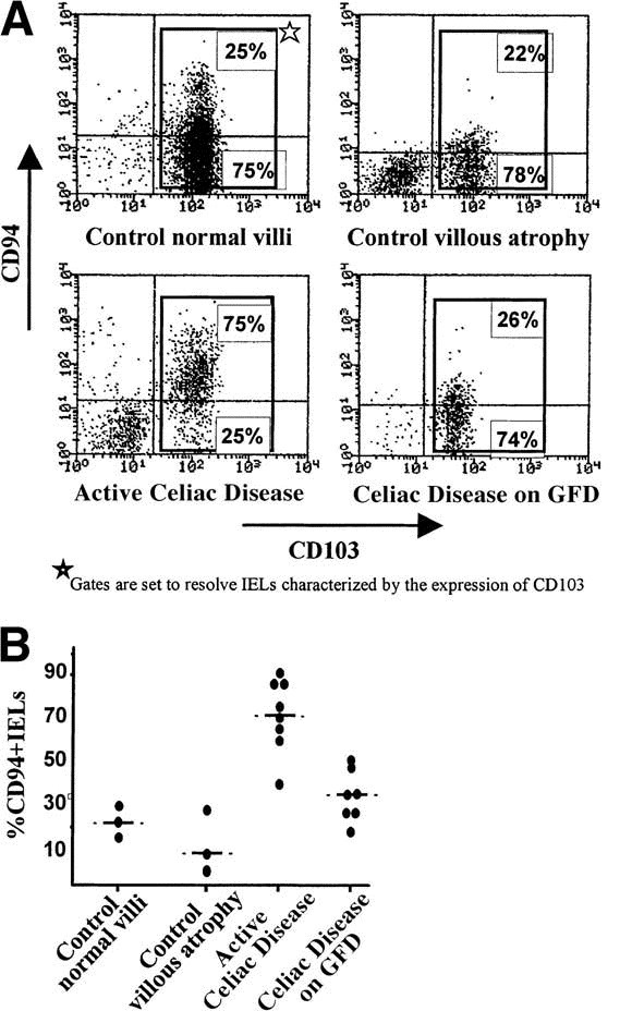
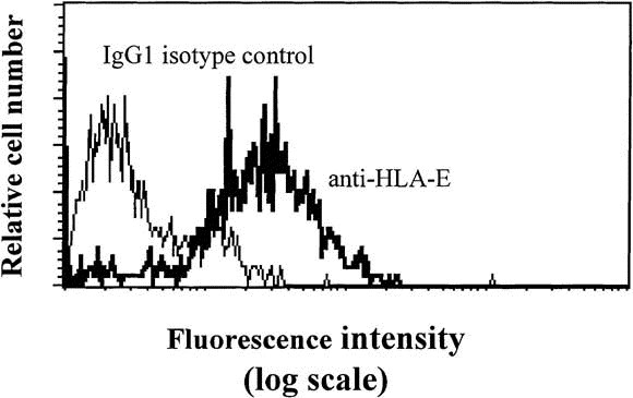
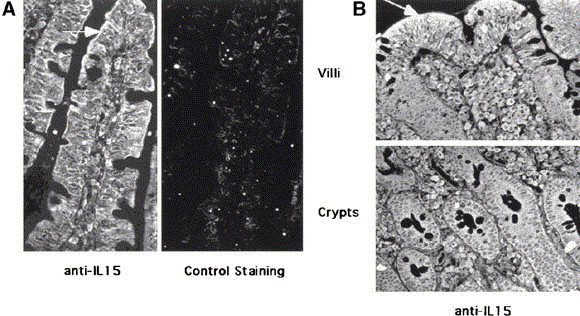
Results: In normal intestine, different proportions of IELs, which were mainly T cells, expressed the NK receptors CD94/NKG2, NKR-P1A, KIR2D/3D, NKp46, Pen5, or CD56. During the active phase of celiac disease, the frequency of CD94(+) IELs, which were mostly alphabeta T cells, was conspicuously increased over controls. In contrast, the expression of other NK markers was not modified. Furthermore, expression of CD94 could be selectively induced in vitro by T-cell receptor activation and/or interleukin 15, a cytokine produced by intestinal epithelial cells.

Conclusions: The gut epithelium favors the development of T cells that express NK receptors. In active celiac disease, there is a specific and selective increase of IELs expressing CD94, the HLA-E-specific NK receptor that may be related to T-cell receptor activation and/or interleukin 15 secretion.


Figures

Fig. 1
Fig. 1
Expression of NK receptors by CD94+ IELs. Flow-cytometric analysis using an anti-CD94 antibody associated with several anti-NK receptor antibodies was performed on normal IELs. The results are shown as percentage (+SD) of CD94+ IELs expressing individual NK receptors.
Fig. 2
Fig. 2
Expansion of CD94+ IELs is celiac disease specific and is associated with disease activity. Immunoperoxidase staining using anti-CD94 antibody was performed on frozen sections of duodenum in (A and B) 45 children and in (C) 38 adults. Results are expressed as numbers of CD94+ IELs per 100 ECs. In both (A) children and (C) adults, the number of CD94+ IELs was significantly increased in active celiac disease (P < 0.001) compared with controls with normal intestine, controls with villous atrophy unrelated to celiac disease, or patients receiving GFD. (B) A longitudinal study in 4 celiac children confirmed that counts of CD94+ IELs returned to normal after GFD.
Fig. 3
Fig. 3
Flow-cytometric analysis of CD94+ IELs in celiac disease. Flow-cytometric analysis using conjugated anti-CD94 and anti-CD103 antibodies was performed on IELs. Expression of the CD103 integrin, characteristic of IELs, was used to distinguish IELs from ECs contaminating the lymphocyte preparation (lower-left quadrant). Results are expressed as percentages of CD94+ and CD94 cells among CD103+ IELs. (A) Representative examples of controls and patients with celiac disease (CD). (B) Summary of the results obtained in 6 controls and 15 celiac patients.
Fig. 4
Fig. 4
Most CD94+ IELs express the TCRαβ receptor. Flow-cytometric analysis of IELs using anti-CD94 vs. anti-TCRαβ or anti-TCRγδ antibodies in a representative patient with untreated celiac disease. Similar results were obtained in all 6 patients studied. Gates are set to resolve CD94+ cells.
Fig. 5
Fig. 5
Expression of HLA-E on KATO EC line. The KATO EC line of gastric origin was stained with 3D12 anti–HLA-E monoclonal antibody or with IgG1 isotype control.
Fig. 6
Fig. 6
IL-15 alone induces the expression of CD94 by IELs but not by PBLs. Altogether, 1 × 10⁵ to 2 × 10⁵ FACS-sorted CD94CD8+ IELs and PBLs were cultured in RPMI plus 10% fetal calf serum alone or in the presence of saturating amounts of IL-15 in round-bottom microwells. For TCR-mediated stimulation, cells were cultured in round-bottom microwells precoated overnight with 10 μg/mL anti-CD3. Cells were analyzed by flow cytometry after 24 hours (IELs) or 48 hours (PBLs) in culture. PBLs are gated on CD8+CD45RO (top row) and CD8+CD45RO+ (middle row), and IELs are gated on CD8+ (bottom row). Quadrants for statistical analysis were set so as to score as negative more than 99% of control-stained cells.
Fig. 7
Fig. 7
Expression of IL-15 protein in celiac intestine. Paraffin sections of duodenal biopsy specimens from (A) a control subject and (B) a patient with active celiac disease were stained with anti–IL-15 monoclonal antibody. For control staining (A), anti–IL-15 antibody was preincubated with recombinant IL-15 at neutralizing concentrations. In controls, IL-15 was mainly detected in the villous ECs. In celiac disease, IL-15 was strongly expressed by both crypt and villous enterocytes as well as by numerous lamina propria cells. Arrows indicate areas of strong positivity for IL-15.

References

    1. Godkin A, Jewell D. The pathogenesis of celiac disease. Gastroenterology. 1998;115:206–210. - PubMed
    1. Sollid LM, Markussen G, Ek J, Gjerde H, Var tdal F, Thorsby E. Evidence for a primary association of celiac disease to a particular HLA-DQ α/β heterodimer. J Exp Med. 1989;169:345–350. - PMC - PubMed
    1. Dieterich W, Ehnis T, Bauer M, Donner P, Volta U, Riecken EO, Schuppan D. Identification of tissue transglutaminase as the autoantigen of celiac disease. Nat Med. 1997;3:797–801. - PubMed
    1. Halstensen TS, Brandtzaeg P. Activated T lymphocytes in the celiac lesion: non-proliferative activation (CD25) of CD4+ α/β cells in the lamina propria but proliferation (Ki-67) of α/β and γ/δ cells in the epithelium. Eur J Immunol. 1993;23:505–510. - PubMed
    1. Kutlu T, Brousse N, Rambaud C, Le Deist F, Schmitz J, Cerf-Bensussan N. Numbers of T cell receptor (TCR) αβ+ but not of TcR γδ+ intraepithelial lymphocytes correlate with the grade of villous atrophy in coeliac patients on a long term normal diet. Gut. 1993;34:208–214. - PMC - PubMed

Publication types

MeSH terms