. 2000 May;66(5):2166-74.
doi: 10.1128/AEM.66.5.2166-2174.2000.

Molecular ecological analysis of the succession and diversity of sulfate-reducing bacteria in the mouse gastrointestinal tract

Affiliations

Molecular ecological analysis of the succession and diversity of sulfate-reducing bacteria in the mouse gastrointestinal tract

B Deplancke et al. Appl Environ Microbiol. 2000 May.

Abstract

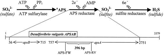
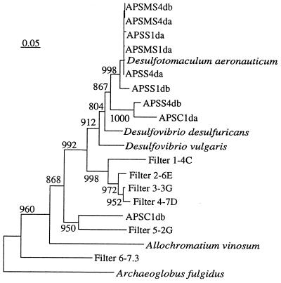
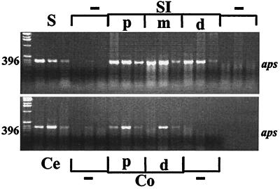
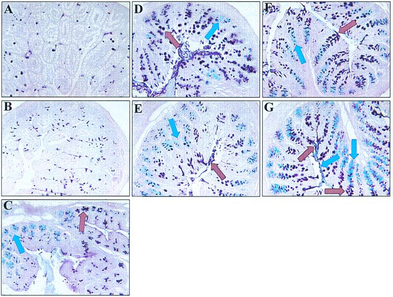
Intestinal sulfate-reducing bacteria (SRB) growth and resultant hydrogen sulfide production may damage the gastrointestinal epithelium and thereby contribute to chronic intestinal disorders. However, the ecology and phylogenetic diversity of intestinal dissimilatory SRB populations are poorly understood, and endogenous or exogenous sources of available sulfate are not well defined. The succession of intestinal SRB was therefore compared in inbred C57BL/6J mice using a PCR-based metabolic molecular ecology (MME) approach that targets a conserved region of subunit A of the adenosine-5'-phosphosulfate (APS) reductase gene. The APS reductase-based MME strategy revealed intestinal SRB in the stomach and small intestine of 1-, 4-, and 7-day-old mice and throughout the gastrointestinal tract of 14-, 21-, 30-, 60-, and 90-day-old mice. Phylogenetic analysis of APS reductase amplicons obtained from the stomach, middle small intestine, and cecum of neonatal mice revealed that Desulfotomaculum spp. may be a predominant SRB group in the neonatal mouse intestine. Dot blot hybridizations with SRB-specific 16S ribosomal DNA (rDNA) probes demonstrated SRB colonization of the cecum and colon pre- and postweaning and colonization of the stomach and small intestine of mature mice only. The 16S rDNA hybridization data further demonstrated that SRB populations were most numerous in intestinal regions harboring sulfomucin-containing goblet cells, regardless of age. Reverse transcriptase PCR analysis demonstrated APS reductase mRNA expression in all intestinal segments of 30-day-old mice, including the stomach. These results demonstrate for the first time widespread colonization of the mouse intestine by dissimilatory SRB and evidence of spatial-specific SRB populations and sulfomucin patterns along the gastrointestinal tract.


Figures

FIG. 1
FIG. 1
Biochemistry and genetics of dissimilatory sulfate reduction. The molecular ecology strategy outlined targets the dissimilatory sulfate reduction pathway and selectively amplifies the APS reductase A subunit gene or corresponding RNA transcripts from composite intestinal DNA or RNA samples, using the primer set APS-FW and APS-RV. The positions of nucleotides are according to the D. vulgaris APS reductase sequence (D. vulgaris APSAB, GenBank accession no. Z69372). The APSAB gene is 3,379 bp long and is compromised of subunit genes A and B, as indicated schematically above. The subunit genes A and B code for the enzyme APS reductase. aAPS, adenosine-5′-phosphosulfate.
FIG. 2
FIG. 2
PCR amplification of the 396-bp APS reductase fragment (aps) from positive control SRB strains (D. ruminis, lane 1; D. thermobenzoicum, lane 2; D. vulgaris, lane 3; D. curvatus, lane 4; and D. salexigens, lane 5) and the presence of APS reductase amplicons of the correct size (396 bp) from a sediment filter DNA sample (lane 6). PCR amplification of DNA from negative control bacteria strains (E. faecalis, lane 7; B. ovatus, lane 8; and E. coli, lane 9) did not yield APS reductase amplicons. M corresponds to a 1-kb ladder (Gibco BRL).
FIG. 3
FIG. 3
Agarose gel showing presence or absence of intestinal SRB in distinct intestinal regions of C57BL/6J mice of different ages (A, 1 day after birth; B, 14 days; C, 21 days; D, 90 days) based on detection of APS reductase amplicons (aps) of the correct size (396 bp). D. vulgaris was used as a positive (+) control, and DNA from mouse kidney as a negative (−) control. Intestinal contents of three mice were analyzed at each sampling point. S, stomach; SI, small intestine; Ce, cecum; Co, colon; p, proximal; m, middle; d, distal.
FIG. 4
FIG. 4
DGGE analysis of APS reductase DNA amplicons comparing the banding patterns from distinct mouse intestinal regions (lane 1, stomach; lanes 2 to 4, proximal, middle, and distal SI; lane 5, cecum; and lanes 6 to 7, proximal and distal colon) with three positive control SRB strains (lane 8, D. desulfuricans [b]; lane 9, D. ruminis [a]; and lane 10, D. curvatus [c]). M corresponds to a synthetic marker comprised of known 16S rDNA sequences varying in GC content (see Materials and Methods).
FIG. 5
FIG. 5
Phylogenetic placement of APS reductase sequences from intestinal and environmental samples. The archaeal sequence (Archaeoglobus fulgidus) was used as the outgroup for rooting the tree. Numbers above each node are confidence levels generated from 1,000 bootstrap trees (8). The scale bar is in fixed nucleotide substitutions per sequence position. Intestinal APS reductase sequences are amplicons from clones (a and b) from the stomach (APSS1da and APSS1db), middle SI (APSMS1da), and cecum (APSC1da and APSC1db) of a 1-day-old mouse and from the stomach (APSMS4da and APSS4db) and middle SI (APSMS4da and APSMS4db) of a 4-day-old mouse. Filters 1-4C, 2-6E, 3-3G, 4-7D, 5-2G, and 6-7.3 represent APS reductase amplicons from six clones from the biofilm of the water filter.
FIG. 6
FIG. 6
Agarose gel comparing the presence and intensity of RT-PCR APS reductase amplicons (aps) in intestinal regions from three 30-day-old mice (stomach [S]; proximal [p], middle [m], and distal [d] SI; cecum [Ce]; and proximal [p] and distal [d] colon [Co]). Samples without reverse transcriptase served as a negative control (−) to screen for DNA contamination. APS reductase mRNA expression was observed in all intestinal regions.
FIG. 7
FIG. 7
Histological analysis of distinct intestinal regions of C57BL/6J mice by means of high iron diamine-alcian blue (pH 2.5) histology to differentiate sialated mucins (blue stain, blue arrow) from sulfated mucins (brown stain, brown arrow) (magnification, ×20). (A and B) Increase of sulfomucin-containing goblet cells in the distal SI before weaning (A, 14-day-old mouse) compared to after weaning (B, 30-day-old mouse). (C) Spatial mucin distribution in the cecum of a 90-day-old mouse. Sialated mucins (blue) predominate in the proximal cecum, while sulfated mucins (brown) predominate in the distal cecum. (D to G) Succession of mucin types in the mouse distal colon (D, 14 day old; E, 30 day old; F, 60 day old; and G, 90 day old).

References

    1. Amann R I, Binder B J, Olsen R J, Chisholm S W, Devereux R, Stahl D A. Combination of 16S rRNA-targeted oligonucleotide probes with flow cytometry for analyzing mixed microbial populations. Appl Environ Microbiol. 1990;56:1919–1925. - PMC - PubMed
    1. Beerens H, Romond C. Sulfate-reducing anaerobic bacteria in human feces. Am J Clin Nutr. 1977;30:1770–1776. - PubMed
    1. Butine T J, Leedle J A. Enumeration of selected anaerobic bacterial groups in cecal and colonic contents of growing-finishing pigs. Appl Environ Microbiol. 1989;55:1112–1116. - PMC - PubMed
    1. Campbell L L, Postgate J R. Classification of the spore forming sulfate-reducing bacteria. Bacteriol Rev. 1965;29:359–363. - PMC - PubMed
    1. Corfield A P, Wagner S A, Clamp J R, Kriaris M S, Hoskins L C. Mucin degradation in the human colon: production of sialidase, sialate O-acetylesterase, N-acetylneuraminate lyase, arylesterase and glycosulfatase activities by strains of faecal bacteria. Infect Immun. 1992;66:3971–3978. - PMC - PubMed

Publication types

MeSH terms

LinkOut - more resources