. 2000 May;38(5):1869-75.
doi: 10.1128/JCM.38.5.1869-1875.2000.

Molecular differentiation of seven Malassezia species

Affiliations

Molecular differentiation of seven Malassezia species

A K Gupta et al. J Clin Microbiol. 2000 May.

Abstract

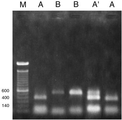
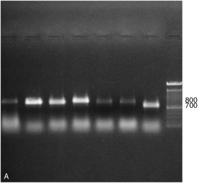
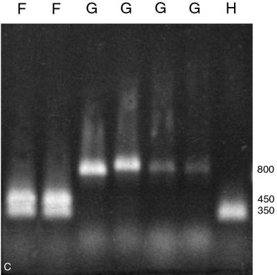
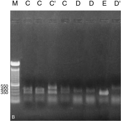
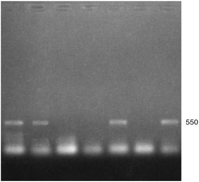
A system based on PCR and restriction endonuclease analysis was developed to distinguish the seven currently recognized Malassezia species. Seventy-eight strains, including authentic culture collection strains and routine clinical isolates, were investigated for variation in the ribosomal DNA repeat units. Two genomic regions, namely, the large subunit of the ribosomal gene and the internal transcribed spacer (ITS) region, were amplified by PCR, and products were digested with restriction endonucleases. The patterns generated were useful in identification of five out of seven Malassezia species. M. sympodialis was readily distinguishable in that its ITS region yielded a 700-bp amplified fragment, whereas the other six species yielded an 800-bp fragment. M. globosa and M. restricta were very similar in the regions studied and could be distinguished only by performing a hot start-touchdown PCR on primers for the beta-tubulin gene. Primers based on the conserved areas of the Candida cylindracea lipase gene, which were used in an attempt to amplify Malassezia lipases, yielded an amplification product after annealing at 55 degrees C only with M. pachydermatis. This specific amplification may facilitate the rapid identification of this organism.


Figures

FIG. 1
FIG. 1
Restriction patterns obtained by AvaI digestion of amplified product from the LSU region (primers 26S-S and 26S-A). A, B, and A′, PCR-REA type; M, 100-bp ladder used as a size marker. Strains loaded in lanes from left to right, JF4 (M. furfur), 99F 160 (M. globosa), ATCC 14521 (M. pachydermatis), CBS 1878 (M. furfur), and 97F 8615 (M. sympodialis). A polaroid picture of the gel was scanned in the computer, and the relevant lanes were aligned together to create this image. Numbers on the left are base pairs.
FIG. 2
FIG. 2
(A) Amplification of seven Malassezia species using primers for the ITS region. Lanes (left to right): M. furfur (CBS 1878), M. pachydermatis (ATCC 14521), M. slooffiae (CBS 7956), M. obtusa (98F 3529), M. globosa (98F 6443), M. restricta (YKM 31), M. sympodialis (CBS 7222), and 100-bp ladder. Numbers on the right are base pairs. (B) Restriction patterns obtained by EcoRI digestion of amplified products from the ITS region (primers ITS 1 and ITS 4). Letters represent PCR-REA types. Lanes (left to right): M. furfur (CBS 1878), M. pachydermatis (ATCC 14521), M. slooffiae (CBS 7956), M. obtusa (98F 3529), M. globosa (98F 6443), M. restricta (YKM 31), M. sympodialis (CBS 7222), and M. sympodialis (GM 323). This image was generated by scanning a polaroid picture of the gel in the computer. Numbers on the left are base pairs. (C) Restriction patterns obtained by NcoI digestion of amplified products from the ITS region (primers ITS 1 and ITS 4). Letters represent PCR-REA types. Lanes (left to right): M. furfur (CBS 1878), M. pachydermatis (ATCC 14521), M. slooffiae (CBS 7956), M. obtusa (98F 3529), M. globosa (98F 6443), M. restricta (YKM 31), and M. sympodialis (CBS 7222). This image was generated by scanning a polaroid picture of the gel in the computer. Numbers on the right are base pairs.
FIG. 2
FIG. 2
(A) Amplification of seven Malassezia species using primers for the ITS region. Lanes (left to right): M. furfur (CBS 1878), M. pachydermatis (ATCC 14521), M. slooffiae (CBS 7956), M. obtusa (98F 3529), M. globosa (98F 6443), M. restricta (YKM 31), M. sympodialis (CBS 7222), and 100-bp ladder. Numbers on the right are base pairs. (B) Restriction patterns obtained by EcoRI digestion of amplified products from the ITS region (primers ITS 1 and ITS 4). Letters represent PCR-REA types. Lanes (left to right): M. furfur (CBS 1878), M. pachydermatis (ATCC 14521), M. slooffiae (CBS 7956), M. obtusa (98F 3529), M. globosa (98F 6443), M. restricta (YKM 31), M. sympodialis (CBS 7222), and M. sympodialis (GM 323). This image was generated by scanning a polaroid picture of the gel in the computer. Numbers on the left are base pairs. (C) Restriction patterns obtained by NcoI digestion of amplified products from the ITS region (primers ITS 1 and ITS 4). Letters represent PCR-REA types. Lanes (left to right): M. furfur (CBS 1878), M. pachydermatis (ATCC 14521), M. slooffiae (CBS 7956), M. obtusa (98F 3529), M. globosa (98F 6443), M. restricta (YKM 31), and M. sympodialis (CBS 7222). This image was generated by scanning a polaroid picture of the gel in the computer. Numbers on the right are base pairs.
FIG. 3
FIG. 3
Hot start-TD PCR amplification product obtained using primers for the β-tubulin gene (12). Lanes (left to right): YKM 31 (M. restricta), CBS 7877 (M. restricta), 98F 9925 (M. sympodialis), 98F-7317 (M. globosa), JF4 (M. furfur); 98F-8316 (M. obtusa), and CBS 7956 (M. slooffiae). Positive amplification of a band of ∼550 bp is seen in lanes 1, 2, 5, and 7.

References

    1. Anaissie E, Bodey G P. Nosocomial fungal infections: old problems and new challenges. Infect Dis Clin N Am. 1989;3:867–882. - PubMed
    1. Anthony R M, Howell S A, Pinters L. Application of DNA typing methods to the study of the epidemiology of Malassezia pachydermatis. Microb Ecol Health Dis. 1994;7:161–168.
    1. Ault G S, Ryschkewitsch C F, Stoner G L. Type-specific amplification of viral DNA using touchdown and hotstart PCR. J Virol Methods. 1994;46:145–156. - PubMed
    1. Belkum A, Boekhout T, Bosboom R. Monitoring spread of Malassezia infections in a neonatal intensive care unit by PCR-mediated genetic typing. J Clin Microbiol. 1994;32:2528–2532. - PMC - PubMed
    1. Boekhout T, Bosboom R W. Karyotyping of Malassezia yeasts: taxonomic and epidemiological implications. Syst Appl Microbiol. 1994;17:147–153.

LinkOut - more resources