. 2000 May;38(5):1953-8.
doi: 10.1128/JCM.38.5.1953-1958.2000.

Rapid identification of Yersinia enterocolitica in blood by the 5' nuclease PCR assay

Affiliations

Rapid identification of Yersinia enterocolitica in blood by the 5' nuclease PCR assay

K Sen. J Clin Microbiol. 2000 May.

Abstract

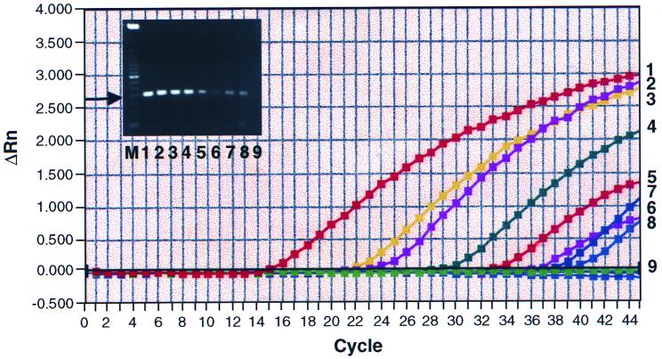
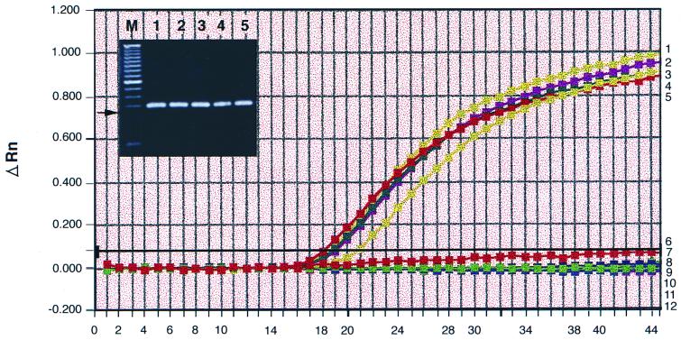
Yersinia enterocolitica accounts for 50% of the clinical sepsis episodes caused by the transfusion of contaminated red blood cells. A 5' nuclease TaqMan PCR assay was developed to detect Y. enterocolitica in blood. Primers and a probe based on the nucleotide sequence of the 16S rRNA gene from Y. enterocolitica were designed. Whole-blood samples were spiked with various numbers of Y. enterocolitica cells, and total chromosomal DNA was extracted. When the TaqMan PCR assay was performed, as few as six bacteria spiked in 200 microliter of blood could be detected. The assay was specific and did not detect other Yersinia species. The TaqMan assay is easy to perform, takes 2 h, and has the potential for use in the rapid detection of Y. enterocolitica contamination in stored blood units.


Figures

FIG. 1
FIG. 1
Detection limit of the TaqMan assay for Y. enterocolitica. Chromosomal DNA from a pure culture of serotype O:3 was isolated as described in Materials and Methods. Quantities of 100 ng, 10 ng, 1 ng, 100 pg, 10 pg, 1 pg, 100 fg, 5 fg, and 1 fg, represented by amplification plots 1 to 9, respectively, were used as template in each 50 μl of PCR mixture. The gel inset shows 5 μl of the corresponding product from each reaction, which was examined on a 2.5% agarose gel. Lane M is the molecular size standard, consisting of a 50-kb DNA ladder. Lanes 1 to 9 correspond to plots 1 to 9. The arrow indicates the position of the 201-bp product obtained as a result of amplification of the region from nucleotides 47 to 247 of the 16S rRNA gene with primers 16SF and 16SR.
FIG. 2
FIG. 2
Specificity of the TaqMan assay for Y. enterocolitica. Chromosomal DNAs (20 ng each) from five Y. enterocolitica serotypes (Y288; O:1,2,3; O:3; O:5,27; and O:20) were used as templates in TaqMan PCR, and their amplifications are represented in plots 1 to 5, respectively. The gel inset shows the 201-bp product from each PCR, and lanes 1 to 5 correspond to plots 1 to 5. The arrow indicates the position of the 201-bp product. Chromosomal DNAs (200 ng each) from seven other bacterial species listed in Table 1 were also tested with primers 16SF and 16SR and probe YE1, and their amplifications are represented in plots 6 to 12. The cycle numbers are on the x axis.
FIG. 3
FIG. 3
Detection limit of the TaqMan assay for Y. enterocolitica in blood samples. Different amounts of Y. enterocolitica cells were spiked into blood, and the total DNA was isolated as described in Materials and Methods. (A) Bacteria were spiked into 200 μl of blood, and DNA was isolated by the QIAamp blood kit in a final volume of 100 μl. The corresponding numbers of cells spiked are as follows: plot 1, none (unseeded blood); plot 2, 30 cells/ml (1.2 cell equivalent/50 μl of PCR mixture); plot 3, 60 cells/ml (2.4 cell equivalent/50 μl of PCR mixture); plot 4, 300 cells/ml; plot 5, 3,000 cells/ml; and plot 6, 30,000 cells/ml. The gel inset shows the products obtained, and lanes 1 to 6 correspond to plots 1 to 6. Lane 7 is a no-template control. (B) Bacteria were spiked into 100 μl of blood, and DNA was extracted by the Dynal DNA Direct kit in a final volume of 75 μl. The corresponding cell numbers are as follows: plot 1, none (unseeded blood); plot 2, 30 cells/ml (1.2 cell equivalent/PCR mixture); plot 3, 300 CFU/ml (12 cell equivalents/PCR mixture); plot 4, 3,000 cells; and plot 5, 30,000 cells. The gel inset represents the products obtained, and lanes 1 to 5 correspond to plots 1 to 5. M is the 50-bp molecular size DNA ladder, and the arrow indicates the 201-bp product.

References

    1. Akane A, Matsubara K, Nakamura H, Takahashi S, Kimura K. Identification of the heme compound copurified with deoxyribonucleic acid (DNA) from bloodstains, a major inhibitor of polymerase chain reaction (PCR) amplification. J Forensic Sci. 1994;39:362–372. - PubMed
    1. Altschul S F, Madden T L, Schaffer A A, Zhang J, Zhang Z, Miller W, Lipman D J. Gapped BLAST and PSI-BLAST: a new generation of protein database search programs. Nucleic Acids Res. 1997;25:3389–3402. - PMC - PubMed
    1. Arduino M J, Bland L A, Tipple M A, Aguero S M, Favero M S, Jarvis W R. Growth and endotoxin production of Yersinia enterocolitica and Enterobacter agglomerans in packed erythrocytes. J Clin Microbiol. 1989;27:1483–1485. - PMC - PubMed
    1. Backman A, Lantz P, Radstrom P, Olcen P. Evaluation of an extended diagnostic PCR assay for detection and verification of the common causes of bacterial meningitis in CSF and other biological samples. Mol Cell Probes. 1999;13:49–60. - PubMed
    1. Bassler H A, Flood S J A, Livak K J, Marmaro J, Knorr R, Batt C A. Use of a fluorogenic probe in a PCR-based assay for the detection of Listeria monocytogenes. Appl Environ Microbiol. 1995;61:3724–3728. - PMC - PubMed

MeSH terms

LinkOut - more resources