. 2000 May 1;524 Pt 3(Pt 3):795-806.
doi: 10.1111/j.1469-7793.2000.00795.x.

Functional coupling between glycolysis and excitation-contraction coupling underlies alternans in cat heart cells

Affiliations

Functional coupling between glycolysis and excitation-contraction coupling underlies alternans in cat heart cells

J Hüser et al. J Physiol. .

Abstract

Electromechanical alternans was characterized in single cat atrial and ventricular myocytes by simultaneous measurements of action potentials, membrane current, cell shortening and changes in intracellular Ca2+ concentration ([Ca2+]i). Using laser scanning confocal fluorescence microscopy, alternans of electrically evoked [Ca2+]i transients revealed marked differences between atrial and ventricular myocytes. In ventricular myocytes, electrically evoked [Ca2+]i transients during alternans were spatially homogeneous. In atrial cells Ca2+ release started at subsarcolemmal peripheral regions and subsequently spread toward the centre of the myocyte. In contrast to ventricular myocytes, in atrial cells propagation of Ca2+ release from the sarcoplasmic reticulum (SR) during the small-amplitude [Ca2+]i transient was incomplete, leading to failures of excitation-contraction (EC) coupling in central regions of the cell. The mechanism underlying alternans was explored by evaluating the trigger signal for SR Ca2+ release (voltage-gated L-type Ca2+ current, ICa,L) and SR Ca2+ load during alternans. Voltage-clamp experiments revealed that peak ICa,L was not affected during alternans when measured simultaneously with changes of cell shortening. The SR Ca2+ content, evaluated by application of caffeine pulses, was identical following the small-amplitude and the large-amplitude [Ca2+]i transient. These results suggest that the primary mechanism responsible for cardiac alternans does not reside in the trigger signal for Ca2+ release and SR Ca2+ load. beta-Adrenergic stimulation with isoproterenol (isoprenaline) reversed electromechanical alternans, suggesting that under conditions of positive cardiac inotropy and enhanced efficiency of EC coupling alternans is less likely to occur. The occurrence of electromechanical alternans could be elicited by impairment of glycolysis. Inhibition of glycolytic flux by application of pyruvate, iodoacetate or beta-hydroxybutyrate induced electromechanical and [Ca2+]i transient alternans in both atrial and ventricular myocytes. The data support the conclusion that in cardiac myocytes alternans is the result of periodic alterations in the gain of EC coupling, i. e. the efficacy of a given trigger signal to release Ca2+ from the SR. It is suggested that the efficiency of EC coupling is locally controlled in the microenvironment of the SR Ca2+ release sites by mechanisms utilizing ATP, produced by glycolytic enzymes closely associated with the release channel.


Figures

Figure 1
Figure 1. Electrical and mechanical alternans in atrial (A) and ventricular (B) myocytes
Action potentials were evoked by 2 ms pulses in the current-clamp mode using the perforated patch technique (see Methods). Mechanical alternans were quantified by measuring cell shortening with a video edge detection method. In both cell types electrical stimulation at frequencies of 0.5–1.5 Hz evoked discordant electrical (top trace) and mechanical (bottom trace) alternans. To the right in A and B two APs recorded during subsequent small (○)- and large (•)-amplitude shortening are superimposed to illustrate the differences in duration and kinetics.
Figure 2
Figure 2. Spatio-temporal characteristics of [Ca2+]i transients during alternans in atrial and ventricular cells
A, alternans (a) in atrial cells is characterized by marked variations in the spatial profile of the [Ca2+]i transients (b). Local [Ca2+]i profiles (c), recorded during large- and small-amplitude [Ca2+]i transients, revealed [Ca2+]i gradients directed from subsarcolemmal (ss) regions toward central regions (ct) of the cell. The small-amplitude [Ca2+]i transients were spatially restricted to the subsarcolemmal regions. B, in ventricular myocytes during alternans (a) the [Ca2+]i transients alternate in a spatially homogeneous pattern (b). During the large or the small transient the rise time and the size of the transients were similar in the subsarcolemmal (ss) and in the centre region (ct) of the cell (c).
Figure 3
Figure 3. ICa, L and cell shortening during alternans
Simultaneous measurements of cell shortening (top) and ICa, L (bottom) in an atrial myocyte. Holding potential VH=−40 mV, test potential = 0 mV. Despite large differences in cell shortening during alternans peak ICa, Lremained constant.
Figure 4
Figure 4. SR Ca2+ content during alternans
SR Ca2+ content was evaluated with 10 mM caffeine pulses in atrial myocytes. Caffeine was applied twice to the same cell. The amplitude of the caffeine-induced [Ca2+]i transient did not reveal any significant differences after large- (left panel) and small-amplitude (right panel) [Ca2+]i transients, indicating that SR Ca2+ content remained constant and did not alternate.
Figure 5
Figure 5. Effect of β-adrenergic stimulation on electromechanical alternans
Discordant electromechanical alternans were elicited in a single ventricular cell by electrical stimulation (1.1 Hz) under current-clamp conditions at room temperature. Top panel: application of isoproterenol (1.5 nM) reversed alternans. Upon wash-out of isoproterenol electromechanical alternans resumed. Bottom panels, action potential recordings and cell shortening at expanded time scale recorded at times indicated by the letters a to e in the top panel.
Figure 6
Figure 6. Alternans of cell shortening, action potentials and ICa, L induced by pyruvate
A, discordant electromechanical alternans elicited by external application of 10 mM pyruvate in a ventricular myocyte under current-clamp conditions. B, mechanical alternans recorded from voltage-clamped atrial myocytes superfused with pyruvate. During alternans ICa, L remained constant. Holding potential VH=−40 mV, test potential = 0 mV.
Figure 7
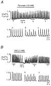
Figure 7. [Ca2+]i transient alternans induced by inhibition of glycolysis
Inhibition of glycolysis by exposure to 10 mM pyruvate (A) and 1 mM iodoacetic acid (IAA; B) caused [Ca2+]i transient alternans in an atrial myocyte. Alternans were reversible upon wash-out of the inhibitors of glycolysis. The bottom traces show the [Ca2+]i transient at expanded time scales.

References

    1. al-Habori M. Microcompartmentation, metabolic channeling and carbohydrate metabolism. International Journal of Biochemistry and Cell Biology. 1995;27:123–132. - PubMed
    1. Badeer HS, Ryo UY, Gassner WF, Kass EJ, Cavaluzzi J, Gilbert JL, Brooks CM. Factors affecting pulsus alternans in the rapidly driven heart and papillary muscle. American Journal of Physiology. 1967;213:1095–1101. - PubMed
    1. Berlin JR. Spatiotemporal changes of Ca2+ during electrically evoked contractions in atrial and ventricular cells. American Journal of Physiology. 1995;269:H1165–1170. - PubMed
    1. Bers DM. Excitation-Contraction Coupling and Cardiac Contractile Force. Boston MA, USA: Kluwer Academic Publishers; 1991.
    1. Blatter LA, Hüser J, Rios E. Sarcoplasmic reticulum Ca2+ release flux underlying Ca2+ sparks in cardiac muscle. Proceedings of the National Academy of Sciences of the USA. 1997;94:4176–4181. - PMC - PubMed

Publication types

MeSH terms

LinkOut - more resources