. 2000 May 2;19(9):1963-73.
doi: 10.1093/emboj/19.9.1963.

MEF2 responds to multiple calcium-regulated signals in the control of skeletal muscle fiber type

Affiliations

MEF2 responds to multiple calcium-regulated signals in the control of skeletal muscle fiber type

H Wu et al. EMBO J. .

Abstract

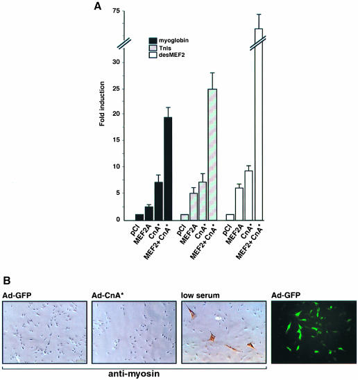
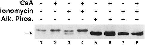
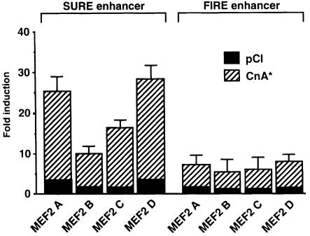
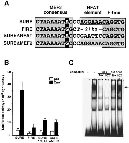
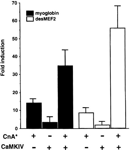
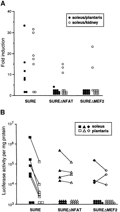
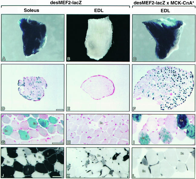
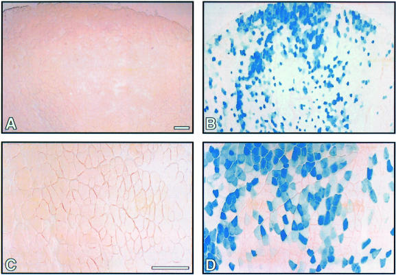
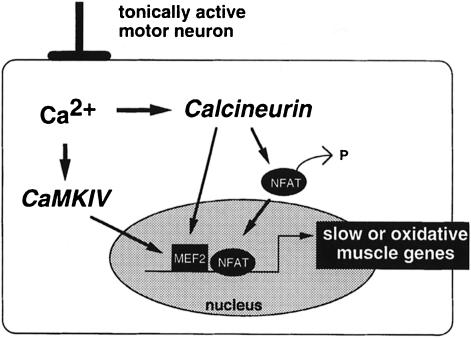
Different patterns of motor nerve activity drive distinctive programs of gene transcription in skeletal muscles, thereby establishing a high degree of metabolic and physiological specialization among myofiber subtypes. Recently, we proposed that the influence of motor nerve activity on skeletal muscle fiber type is transduced to the relevant genes by calcineurin, which controls the functional activity of NFAT (nuclear family of activated T cell) proteins. Here we demonstrate that calcineurin-dependent gene regulation in skeletal myocytes is mediated also by MEF2 transcription factors, and is integrated with additional calcium-regulated signaling inputs, specifically calmodulin-dependent protein kinase activity. In skeletal muscles of transgenic mice, both NFAT and MEF2 binding sites are necessary for properly regulated function of a slow fiber-specific enhancer, and either forced expression of activated calcineurin or motor nerve stimulation up-regulates a MEF2-dependent reporter gene. These results provide new insights into the molecular mechanisms by which specialized characteristics of skeletal myofiber subtypes are established and maintained.


Figures

None
Fig. 1. The transactivating function of MEF2 is stimulated by calcineurin in C2C12 myogenic cells. (A) Luciferase reporter constructs were prepared using promoter/enhancer elements from the myoglobin or TnI genes, or by three copies of a high affinity MEF2 binding site from the desmin promoter (desMEF2). Expression of luciferase driven by each of these reporter plasmids was determined following co-transfection of expression plasmids containing either no insert (pCI) or cDNAs encoding MEF2A or CnA*. Data are expressed relative to the luciferase activity observed in the control state (pCI co-transfection) and represent mean values (± SEM) from five or more independent experiments. All results are corrected for variations in transfection efficiency by normalization to expression of a co-transfected pCMV-lacZ plasmid. (B) The effect of calcineurin to up-regulate myoglobin and TnIs enhancer function is not attributable to a generalized stimulation of muscle differentiation. The three leftward panels show immunostains using anti-myosin antibody of C2C12 myoblasts infected with an adenoviral vector expressing CnA* (Ad-CnA*), or green fluorescent protein (Ad-GFP), or grown in low serum to promote differentiation. Adenovirus-infected cells are identified by expression of GFP (right panel).
None
Fig. 2. DNA binding of MEF2 following activation by calcineurin. C2C12 cells were infected with recombinant adenoviral vectors encoding either green fluorescent protein (Ad-GFP) or CnA* (Ad-CnA*). (A) Luciferase reporter gene expression following transfection of the desMEF2 enhancer construct. Data are presented as in Figure 1 and represent mean values of two independent experiments. (B) Electrophoretic mobility shift assays (EMSA) of nuclear proteins from adenovirus-infected cells using a high affinity MEF2 binding site as the labeled oligonucleotide probe. The identity of the MEF2–DNA complex (arrow) is confirmed by the change in mobility in the presence of anti-MEF2 antibodies, and by competition with unlabeled MEF2 binding oligonucleotide (competitor). In contrast to the clear functional effect on transcription, no detectable differences in DNA binding by MEF2 are observed in the presence of activated calcineurin. Pre, preimmune serum.
None
Fig. 3. Calcineurin-dependent dephosphorylation of MEF2. Proteins extracted from C2C12 myotubes were separated by SDS–PAGE and immunoblots were probed with anti-MEF2 antibody. A hypophosphorylated form of MEF2A (arrow) is evident in cells exposed to a calcium ionophore (1 µM ionomycin, lane 3). Dephosphorylation of MEF2A in the presence of ionomycin was inhibited by the calcineurin antagonist cyclosporin A (250 nM CsA, lane 4). The identity of the more rapidly migrating band as a hypophosphorylated form of MEF2A was confirmed by incubation of protein extracts with alkaline phosphatase (Alk. Phos., lanes 5–8). Similar findings were observed in each of three independent experiments.
None
Fig. 4. All MEF2 isoforms enhance calcineurin-dependent transactivation of a SURE enhancer. Expression plasmids containing cDNAs encoding each of the four isoforms of MEF2 were cotransfected with luciferase reporter plasmids controlled by either the SURE or FIRE enhancers (Nakayama et al., 1996). Luciferase activity following forced expression of each MEF2 isoform was measured in the absence (solid bars, pCI) or presence (dashed bars, CnA*) of activated calcineurin. Fold induction is relative to enhancer activity without cotransfected MEF2 or calcineurin. Data are presented as described in Figure 1.
None
Fig. 5. MEF2 binding to slow and fast fiber-specific enhancers. (A) Nucleotide sequence alignment of segments of the rat TnI SURE and quail TnI FIRE enhancers (Nakayama et al., 1996) that include MEF2, NFAT and E box motifs. The putative MEF2 binding site in FIRE has a G nucleotide rather than an A nucleotide in position 9 of the MEF2 consensus binding motif (darkly shaded), and an NFAT consensus binding motif (boxed) found in SURE is not present in FIRE. The sequences of two variant forms of the SURE enhancer generated by site-directed mutagenesis are also illustrated. SUREΔNFAT and SUREΔMEF2 refer to variants in which NFAT and MEF2 binding motifs, respectively, are altered while leaving the remainder of the SURE enhancer sequence intact. The SUREΔMEF2 includes only the indicated single nucleotide substitution within an otherwise intact SURE enhancer. (B) Luciferase reporter constructs were prepared using the SURE or FIRE enhancers, or the indicated mutants thereof. Expression of luciferase following transfection of each of these reporter plasmids was assessed in the presence or absence of CnA*. Data are presented as in Figure 1 and represent mean values of two to four independent experiments. (C) EMSA of nuclear proteins from C2C12 cells using a high affinity MEF2 binding site (MCK-MEF2) as the labeled oligonucleotide probe. Unlabeled oligonucleotides (competitor) representing either the probe sequence (MCK-MEF2) or the MEF2 binding site from the SURE enhancer compete effectively with the labeled probe for binding MEF2, while the MEF2 motif from the FIRE enhancer competes less avidly. The identity of the MEF2–DNA complex (arrow) was confirmed by the change in mobility in the presence of anti-MEF2 antibodies (see Figure 2).
None
Fig. 6. Calcineurin and CaMKIV act synergistically to enhance the transactivating function of MEF2. Luciferase reporter plasmids controlled by the myoglobin or desMEF2 enhancers were cotransfected into C2C12 cells with expression plasmids encoding either constitutively active calcineurin A subunit or CaMKIV. Data are presented as in Figure 1, and represent results of three or more independent experiments.
None
Fig. 7. Slow fiber-specific activity of the SURE enhancer requires intact NFAT and MEF2 binding motifs. Transgenic mice were generated using luciferase reporter constructs controlled by either the native SURE enhancer, or by two variant forms (SUREΔNFAT or SUREΔMEF2; see Figure 5A). Individual transgenic offspring, each representing a different chromosomal insertion event, were examined at 7 weeks of age for luciferase activity in soleus and plantaris muscles, which are enriched in slow, oxidative or fast, glycolytic myofibers respectively, and in kidney. (A) Data are presented as the ratio of transgene expression in soleus versus plantaris muscles (filled circles) as a measure of slow fiber-specific transcription, and as the ratio of transgene expression in soleus muscle versus kidney (open circles) as a measure of muscle-specific transcription. Each point corresponds to an individual transgenic animal. (B) Data are presented as the log of luciferase activity in the soleus (filled symbols) and plantaris (open symbols) skeletal muscles. Each point corresponds to an individual transgenic animal, and lines connect data from the same animal.
None
Fig. 8. The transactivating function of MEF2 is detected selectively in soleus muscles of transgenic mice, and is up-regulated by activated calcineurin. Soleus and EDL muscles were dissected from the hindlimbs of desMEF2-lacZ transgenic mice, or from doubly transgenic mice carrying both the desMEF-lacZ transgene and an MCK-CnA* transgene (see Material and methods), and stained for β-galactosidase activity. The gross appearance of the muscles (A–C), and photomicrographs of serial cross sections stained for β-galactosidase activity (D–I) or myosin ATPase activity (J–L) are shown. The distribution of β-galactosidase appeared to be uniform along the length of positively staining fibers (not shown). (D–F) Bar, 200 µm; (G–L) bar, 40 µm.
None
Fig. 9. The transactivating function of MEF2 is up-regulated by electrical stimulation of the motor nerve. Gastrocnemius muscles were dissected from the hindlimbs of desMEF2-lacZ transgenic mice that were subjected to 5 days of motor nerve stimulation (see Materials and methods). Photomicrographs of muscle cross sections stained for β-galactosidase activity representing the unstimulated contralateral limb (A and C) and the stimulated limb (B and D) of a single animal are shown at two different magnifications (bar, 100 µm). Similar results were observed in each of three animals subject to this stimulation protocol.
None
Fig. 10. A revised mechanistic model for control of slow or oxidative fiber-specific gene expression by motor nerve activity. Findings presented here extend the model we proposed previously (Chin et al., 1998) to emphasize the importance of MEF2 as a downstream effector of calcium-dependent changes in gene expression provoked by a tonically active pattern of motor neuron firing. Signals generated by calcineurin augment the transactivating function of MEF2 in a manner that also is increased by concomitant activation of CaMKIV, and by synergistic interactions with NFAT.

References

    1. Bito H., Deisseroth,K. and Tsien,R.W. (1996) CREB phosphorylation and dephosphorylation: a Ca2+- and stimulus duration-dependent switch for hippocampal gene expression. Cell, 87, 1203–1214. - PubMed
    1. Black B.L. and Olson,E.N. (1998) Transcriptional control of muscle development by myocyte enhancer factor-2 (MEF2) proteins. Annu. Rev. Cell Dev. Biol., 14, 167–196. - PubMed
    1. Black B.L., Martin,J.F. and Olson,E.N. (1995) The mouse MRF4 promoter is trans-activated directly and indirectly by muscle-specific transcription factors. J. Biol. Chem., 270, 2889–2892. - PubMed
    1. Blagden C.S., Currie,P.D., Ingham,P.W. and Hughes,S.M. (1997) Notochord induction of zebrafish slow muscle mediated by Sonic hedgehog. Genes Dev., 11, 2163–2175. - PMC - PubMed
    1. Calvo S., Venepally,P., Cheng,J. and Buonanno,A. (1999) Fiber-type-specific transcription of the troponin I slow gene is regulated by multiple elements. Mol. Cell. Biol., 19, 515–525. - PMC - PubMed

Publication types

MeSH terms