. 2000 May 2;19(9):2024-33.
doi: 10.1093/emboj/19.9.2024.

The tight junction protein ZO-1 and an interacting transcription factor regulate ErbB-2 expression

Affiliations

The tight junction protein ZO-1 and an interacting transcription factor regulate ErbB-2 expression

M S Balda et al. EMBO J. .

Abstract

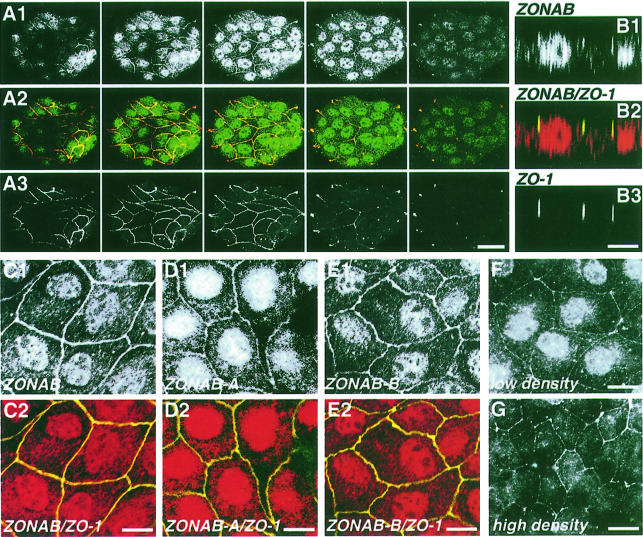
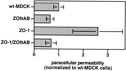
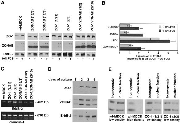
Epithelial tight junctions regulate paracellular diffusion and restrict the intermixing of apical and basolateral plasma membrane components. We now identify a Y-box transcription factor, ZONAB (ZO-1-associated nucleic acid-binding protein), that binds to the SH3 domain of ZO-1, a submembrane protein of tight junctions. ZONAB localizes to the nucleus and at tight junctions, and binds to sequences of specific promoters containing an inverted CCAAT box. In reporter assays, ZONAB and ZO-1 functionally interact in the regulation of the ErbB-2 promoter in a cell density-dependent manner. In stably transfected overexpressing cells, ZO-1 and ZONAB control expression of endogenous ErbB-2 and function in the regulation of paracellular permeability. These data indicate that tight junctions directly participate in the control of gene expression and suggest that they function in the regulation of epithelial cell differentiation.


Figures

None
Fig. 1. Identification of ZONAB. (A) Induced cultures of clones isolated from expression libraries were extracted, fractionated by SDS–PAGE and transferred to nitrocellulose. The membrane was probed with 32P-labeled GST–PDZ3–SH3 fusion protein and exposed to X-ray film. Shown are lanes corresponding to the original clone positive in the expression screening (lane 1), a subclone derived from it (lane 2) and a negative clone expressing an unrelated fusion protein (lane 3). (B) The cDNA sequence of ZONAB-A and the amino acid sequence of the open reading frame. The in-frame upstream stop codon is printed in italic (DDBJ/EMBL/GenBank accession No. AF171061). (C) The cDNA and corresponding amino acid sequence of an additional domain found in some ZONAB transcripts by RT–PCR (DDBJ/EMBL/GenBank accession No. AF171062). (D) Low confluent MDCK cells were grown for 2 days of culture, and expression of ZONAB was detected by immunoblotting with an affinity-purified polyclonal antibody against the C-terminus (lanes 1–3), ZONAB-A (lane 4) or ZONAB-B (lane 5) (wt, wild-type MDCK cells; T:ZONAB-A and -B, MDCK cells stably overexpressing ZONAB-A or -B). (E) The domain structures of ZONAB-A and -B. The domains are also marked in (A). N-PD, N-terminal proline-rich domain; CSD, cold-shock domain; AD, alternative domain; RP-CD, arginine- and proline-rich conserved domain; C-PD, C-terminal proline-rich domain.
None
Fig. 2. Interaction between ZO-1 and ZONAB in wild-type MDCK cells. (A) Cleared lysates of MDCK cells that were at a confluency of ∼20 or 80% were loaded on Sepharose beads with covalently conjugated antibody R40.76 against ZO-1, anti-GST–ZONAB antibody or no antibody (----). The immunoprecipitates were analyzed by immunoblotting with antibodies against ZO-1 and ZONAB (anti-C-terminus), or with secondary antibody only (----). (B) Purified GST fusion proteins containing either the third PDZ domain of ZO-1 (GST–PDZ3), the third PDZ and the SH3 domain (GST–PDZ3–SH3) or the SH3 domain only (GST–SH3) were bound to glutathione–agarose and incubated with diluted recombinant histidine-tagged ZONAB-A. Pull-down of ZONAB-A was tested by immunoblotting. The scheme illustrates the domain structure of ZO-1 with the three PDZ domains, the SH3 domain and the guanylate kinase homology domain (GUK).
None
Fig. 3. Subcellular distribution of ZONAB in subconfluent wild-type and transfected MDCK cells. Wild-type MDCK cells (A–F, low density; G, high density) were fixed and permeabilized with Triton X-100/ethanol. (A) Serial confocal sections taken at 1 µm intervals along the apical–basal axis of an island of cells stained with anti-GST–ZONAB (A1) and anti-ZO-1 (A3) antibodies (bar, 40 µm). (B) Confocal xz-sections of cells stained with the anti-N-terminus ZONAB (B1) and anti-ZO-1 (B3) antibodies. Confocal sections of cells stained with the anti-N-terminus ZONAB (C), anti-ZONAB-A (D) or anti-ZONAB-B (E) antibody; also shown are overlays with ZO-1 staining (C2, D2 and E2) (bars, 10 µm). (F and G) Epifluorescence images of samples labeled with the anti-GST–ZONAB antibody. Note that cells grown to a high density are taller; hence, the focus has been set to the basal end of the junctions to have the nucleus in the same focal plane. This causes the irregular appearance of the junctional staining (bar, 10 µm). Similar ZONAB and ZO-1 staining patterns were observed in cells fixed with methanol and in stable cell lines overexpressing ZONAB.
None
Fig. 4. Regulation of paracellular permeability by ZO-1 and ZONAB. Wild-type and stably transfected MDCK cells overexpressingZONAB-A, ZO-1 or both proteins were plated on filters. TER was measured every day and was not significantly affected by transfection of the two proteins (not shown). After 8 days, the cells were incubated overnight with sodium butyrate to induce higher expression levels of the transfected proteins; this also did not result in differences in TER (not shown). Paracellular flux of [3H]mannitol was then measured during 3 h and normalized to the values obtained from wild-type MDCK cells. Shown are means ± 1 SD of two experiments with four clones overexpressing ZO-1, two clones overexpressing ZONAB, and six clones stably overexpressing both proteins. The absolute numbers were 582 ± 116 c.p.m. for wild-type cells, 378 ± 87 c.p.m. for ZONAB-overexpressing cells, 1513 ± 582 c.p.m. for ZO-1-overexpressing cells and 401 ± 122 c.p.m. for the double transfectants.
None
Fig. 5. ZONAB interacts with specific promoter sequences in vitro. (A) Phosphorylated double-stranded oligonucleotides derived from the ErbB-2 promoter were incubated with different concentrations of GST or GST–ZONAB (2.0, 1.0 and 0.5 µg) in the absence or presence of increasing amounts of unlabeled double-stranded oligonucleotides derived from the ErbB-2 or MDR-1 promoter. Complex formation was analyzed by non-denaturing gel electrophoresis and autoradiography. (B) Binding of ZONAB to inverted CCAAT boxes with different flanking sequences was assayed by direct binding and by the ability to compete for binding of phosphorylated ErbB-2 double-stranded oligonucleotides. The sequences are derived from DDBJ/EMBL/GenBank entries of the genes for ErbB-2 (J05264), p21WAF1/CIP1 (U50603), p27KIP1 (U77914), proliferating cell nuclear antigen (J05614; PCNA), thymidine kinase (M13643; TK-A and TK-B), MDR-1 (L07624), DNA polymerase α (M64481; DPOL), 70 kDa heat-shock protein (M19865; hsp70) and topoisomerase IIa (X66794; TopoII).
None
Fig. 6. Regulation of the ErbB-2 promoter by ZO-1 and ZONAB. Low confluent (A, C, E and G) and confluent (B and F) MDCK cells were co-transfected with a plasmid carrying a fragment of the human ErbB-2 promoter driving the expression of firefly luciferase and a control plasmid carrying a CMV promoter driving the expression of renilla luciferase. (AC) the luciferase reporter plasmid contained the entire 3.6 kb ErbB-2 promoter fragment. (DG) The reporter plasmids contained a shorter fragment of the ErbB-2 promoter with a functional (D: ErbB-2: S) or mutated ZONAB-binding site (D: ErbB-2: S-D, deleted; ErbB-2: S-A, substitution of the inverted CCAAT box). The reporter plasmids were transfected together with empty pCB6 and/or the indicated amounts of vectors resulting in the expression of ZO-1, ZONAB-A, antisense ZONAB RNA (ZONABas), an HA-tagged fragment of ZO-1 containing the third PDZ and the SH3 domain (HA-PDZ3–SH3), or the SH3 domain only (HA-SH3). Empty pCB6 was used to adjust the total DNA concentrations to the same value in all samples. Expression of the luciferases was assayed with a dual-luciferase assay system. (A–C) The ratios obtained (firefly luciferase divided by renilla luciferase) are expressed as a percentage of those obtained by co-transfecting empty pCB6. (D–F) Stimulation or inhibition, respectively, was calculated with respect to control transfections performed with each reporter plasmid in the presence of empty pCB6. The values represent means ± 1 SD of at least two independent experiments performed in duplicate.
None
Fig. 7. Regulation of endogenous ErbB-2 expression by ZO-1 and ZONAB. (A) Wild-type and stably transfected MDCK cells overexpressing ZO-1 and/or ZONAB were synchronized by serum starvation and then collected directly or incubated for 24 h with 10% FCS. Equal amounts of protein were loaded on SDS–gels, and expression of ZO-1, ZONAB and ErbB-2 was quantified by immunoblotting and densitometric scanning. The shown film of the immunoblot with anti-ZONAB antibody is derived from a short exposure to avoid overexposure of lanes derived from cell lines overexpressing ZONAB-A. After longer exposure times, both isoforms of ZONAB were detected in serum-depleted cells without overexpression. (B) The levels of ErbB-2 expression were normalized to the expression in wild-type MDCK cells after serum starvation (three clones of each type of transfection were analyzed in at least two independent experiments). (C) The mRNA levels of ErbB-2 and claudin-4 in cells synchronized by serum starvation were determined by RT–PCR. (D) Total extracts of MDCK cells cultured for 1, 2, 3 and 6 days were separated by SDS–PAGE (equal amounts of protein were loaded in each lane), transferred to nitrocellulose and the expression levels of ZO-1, ZONAB and ErbB-2 were determined by immunoblotting. Cells cultured for 6 days reached full confluency. (E) Nuclear fractions were isolated from wild-type and ZO-1-overexpressing MDCK cells grown to low or high densities. The levels of ZONAB in homogenates and nuclear fractions were determined by immunoblotting.

Similar articles

Cited by

References

    1. Alroy I. and Yarden,Y. (1997) The ErbB signaling network in embryogenesis and oncogenesis: signal diversification through combinatorial ligand–receptor interactions. FEBS Lett., 410, 83–86. - PubMed
    1. Anderson J.M., Stevenson,B.R., Jesaitis,L.A., Goodenough,D.A. and Mooseker,M.S. (1988) Characterization of ZO-1, a protein component of the tight junction from mouse liver and Madin–Darby canine kidney cells. J. Cell Biol., 106, 1141–1149. - PMC - PubMed
    1. Balda M.S. and Anderson,J.M. (1993) Two classes of tight junctions are revealed by ZO-1 isoforms. Am. J. Physiol., 264, C918–C924. - PubMed
    1. Balda M.S. and Matter,K. (1998) Tight junctions. J. Cell Sci., 111, 541–547. - PubMed
    1. Balda M.S., Whitney,J.A., Flores,C., González,S., Cereijido,M. and Matter,K. (1996a) Functional dissociation of paracellular permeability and transepithelial electrical resistance and disruption of the apical–basolateral intramembrane diffusion barrier by expression of a mutant tight junction membrane protein. J. Cell Biol., 134, 1031–1049. - PMC - PubMed

Publication types

MeSH terms

Associated data