. 2000 May 2;19(9):2046-55.
doi: 10.1093/emboj/19.9.2046.

GATA-dependent recruitment of MEF2 proteins to target promoters

Affiliations

GATA-dependent recruitment of MEF2 proteins to target promoters

S Morin et al. EMBO J. .

Abstract

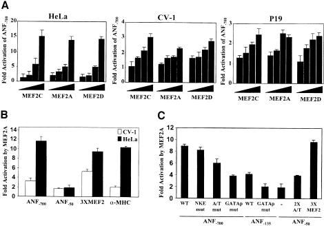
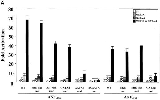
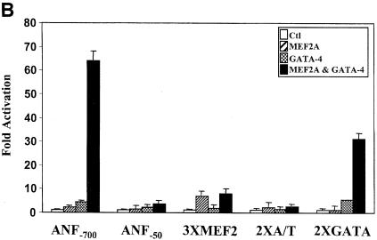
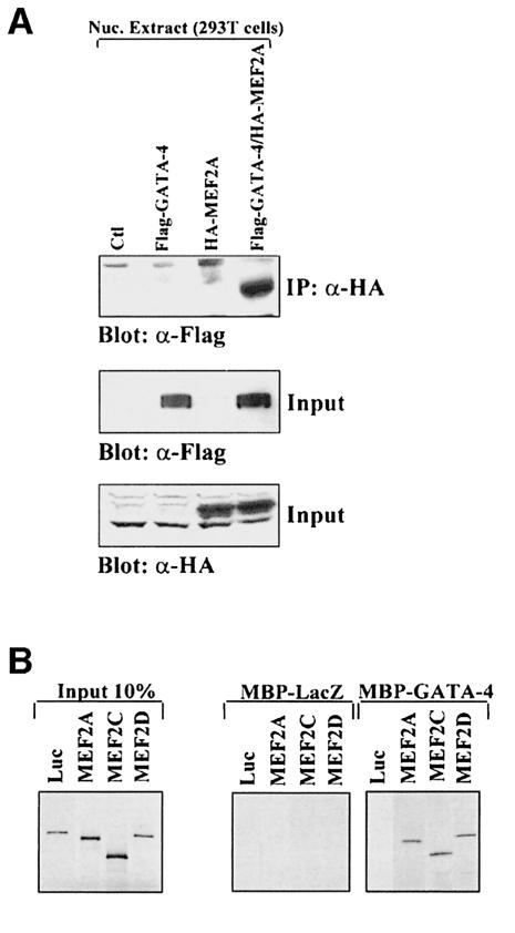
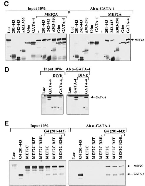
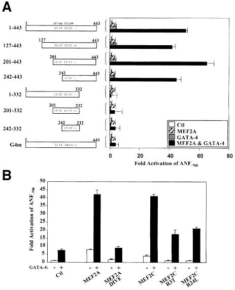
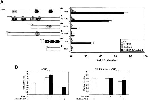
The myocyte enhancer factor-2 (MEF2) proteins are MADS-box transcription factors that are essential for differentiation of all muscle lineages but their mechanisms of action remain largely undefined. In mammals, the earliest site of MEF2 expression is the heart where the MEF2C isoform is detectable as early as embryonic day 7.5. Inactivation of the MEF2C gene causes cardiac developmental arrest and severe downregulation of a number of cardiac markers including atrial natriuretic factor (ANF). However, most of these promoters contain no or low affinity MEF2 binding sites and they are not significantly activated by any MEF2 proteins in heterologous cells suggesting a dependence on a cardiac-enriched cofactor for MEF2 action. We provide evidence that MEF2 proteins are recruited to target promoters by the cell-specific GATA transcription factors, and that MEF2 potentiates the transcriptional activity of this family of tissue-restricted zinc finger proteins. Functional MEF2/GATA-4 synergy involves physical interaction between the MEF2 DNA-binding domain and the carboxy zinc finger of GATA-4 and requires the activation domains of both proteins. However, neither MEF2 binding sites nor MEF2 DNA binding capacity are required for transcriptional synergy. The results unravel a novel pathway for transcriptional regulation by MEF2 and provide a molecular paradigm for elucidating the mechanisms of action of MEF2 in muscle and non-muscle cells.


Figures

None
Fig. 1. The ANF promoter harbors a low-affinity MEF2-binding site. (A) Schematic representation of the ANF promoter. Regulatory elements are boxed and their location relative to the transcription start site is indicated. All these elements are evolutionarily conserved on the ANF promoter from many species. SRE-like is a low-affinity serum response element; the GATAd and GATAp are the distal and proximal GATA-binding sites, respectively. The consensus MEF2-binding site is also shown. The A/T-rich mut sequence indicates the mutations introduced to abolish the A/T-rich element. rANF and hANF are the rat and human ANF promoter, respectively. (B) The A/T-rich element is a low-affinity MEF2-binding site. EMSAs were performed on the MEF2 element of the MCK promoter (MEF2-MCK, left panel) or the A/T-rich element of ANF (A/T-rich, right panel) using in vitro translated MEF2A. In the left panel, the MEF2A binding was competed with different unlabeled ANF probes described in Materials and methods. Only the A/T-rich element of the ANF promoter was able to compete the MEF2A binding. Similar results were obtained with in vitro translated MEF2C and MEF2D.
None
Fig. 2. The low-affinity A/T-rich and the proximal GATA elements contribute to MEF2-dependent ANF promoter activation. (A) Dose-dependent ANF–700 promoter activation by MEF2A, MEF2C and MEF2D in HeLa, CV-1 and P19 cell lines. Transient transfections were performed using 50 ng, 100 ng, 500 ng and 1 µg of MEF2 expression vector. Note the fold-activation difference between HeLa and CV-1 or P19 cells. (B) Preferential activation of the ANF–700 and αMHC promoters, but not an artificial MEF2 reporter, in HeLa cells. Transfections were performed using 1 µg of MEF2A expression vector. Similar results were obtained using MEF2C and MEF2D. (C) The low-affinity A/T-rich and the proximal GATA elements contribute to MEF2-dependent ANF promoter activation. Transfections were performed in HeLa cells using 1 µg of MEF2A expression vector. Similar results were obtained using MEF2C and MEF2D. 3XMEF2 and 2XA/T are the MEF2-MCK and the ANF A/T-rich elements trimerized and dimerized, respectively, in front of the ANF–50 minimal promoter.
None
Fig. 3. The MEF2 and GATA transcription factors cooperatively activate the ANF–700 promoter. (A) MEF2A, MEF2C and MEF2D functionally interact with GATA-4. Cotransfections were performed in HeLa cells using the ANF-luc–700 construct and 1 µg of MEF2A, MEF2C or MEF2D expression vector in the absence (–) or presence (+) of 1.5 µg of GATA-4 expression vector. (B) MEF2 proteins functionally interact with a subset of GATA proteins. Cotransfections were performed as in (A) using 1.5 µg of various GATA expression vectors in the absence (–) or presence (+) of 1 µg of MEF2C expression vector. Note that cooperative interaction between MEF2A and the different GATA factors was identical to the one shown here for MEF2C and GATA-1 to -6. Similar results were also obtained in the CV1 cell line.
None
Fig. 4. The proximal GATA element is necessary and sufficient for MEF2–GATA synergy. Cotransfections were performed in HeLa cells using various promoter constructs and 1 µg of MEF2A and/or 1.5 µg of GATA-4 expression vectors. The ANF promoter constructs used are described in Materials and methods. 3XMEF2 and 2XA/T are the MEF2-MCK and the ANF A/T-rich elements trimerized and dimerized, respectively, in front of the ANF–50 minimal promoter. 2XGATA is a dimer of the BNP GATA elements in front of the minimal BNP promoter.
None
Fig. 5. MEF2 proteins physically interact with GATA-4. (A) MEF2A interacts in vivo with GATA-4. Nuclear extracts from 293T cells transfected with empty vectors (Ctl), Flag-GATA-4 and/or HA-MEF2A were immunoprecipitated using an anti-HA antibody, separated on 10% SDS–PAGE, transferred to PVDF membranes, and subjected to immunoblotting using an anti-Flag antibody (top panel). The lower two panels are Western blots carried out on the same nuclear extracts using either HA (to reveal tagged MEF2A proteins) or Flag (to reveal tagged GATA-4 proteins) antibodies. (B) MEF2A proteins interact in vitro with GATA-4. Pull-down assays were performed using immobilized, bacterially produced MBP fusions (MBP-GATA-4 and MBP-LacZ as control) and in vitro translated 35S-labeled MEF2A, MEF2C, MEF2D or luciferase (luc) protein. The protein complexes were resolved on 10% SDS–PAGE. (C) The physical interaction between GATA-4 and MEF2 requires the C-terminal zinc finger DNA-binding domain of GATA-4. Full-length GATA-4 and various GATA-4 mutants (depicted in Figure 6A) were in vitro cotranslated with MEF2A and co-immunoprecipitated using an antibody directed against the extreme C-terminus of GATA-4. The protein complexes were resolved on 15% SDS–PAGE. The asterisks highlight GATA protein bands. (D) The DNA-binding domain of MEF2 is sufficient for interaction with GATA-4. MEF2A DIVE (aa 1–86) retains the MADS and MEF2 domains. Co-immunoprecipitations were performed as described in (C). The asterisks highlight the MEF2A DIVE band. The protein complexes were resolved on 20% SDS–PAGE. (E) MEF2 DNA-binding-defective mutants interact with GATA-4. MEF2C R3T and MEF2C R24L do not bind DNA but are still able to dimerize. A deleted GATA-4 construct [G4 (201–443)] was used to differentiate between GATA-4 and MEF2C, which have similar electrophoretic mobility. Co-immunoprecipitations were performed as described in (C). The protein complexes were resolved on 10% SDS–PAGE.
None
Fig. 6. Mapping of the GATA-4 and MEF2 domains required for synergy. (A) The C-terminal activation domain of GATA-4 is required for MEF2 synergy. Cotransfections were performed in HeLa cells on the ANF–700 promoter construct using 1 µg of MEF2 and 1.5 µg of GATA-4 expression vectors. (B) The C-terminal activation domain of MEF2, but not its DNA-binding capacity, is required for synergy with GATA-4, as shown by the ability of MEF2C R3T and MEF2C R24L to synergize with GATA-4. Note that the DNA-binding domain (MEF2A DIVE) is not sufficient to support functional synergy although it interacts physically with GATA-4 as shown in the previous figure.
None
Fig. 7. The MEF2–GATA-4 synergy: a mechanism for MEF2 action in the heart. (A) The MEF2–GATA-4 synergy is not limited to the ANF promoter. HeLa cells were cotransfected with 1 µg of MEF2A and 1.5 µg of GATA-4 expression vectors together with various cardiac promoters. Except for the cardiac α-actin promoter that was from chicken, all other promoters used are from rat and are described in Materials and methods. TK81 is the thymidine kinase –81 bp promoter. Elements shaded in black and gray are high- and low-affinity sites, respectively, as determined by DNA-binding assays. (B) A dominant-negative MEF2 protein decreases ANF promoter activity in cardiomyocytes. Primary culture of cardiomyocytes was transfected with the wild-type ANF–135 (left panel) or GATA-mutated ANF–135 promoter (GATAp mut/ANF–135, right panel) and no (–), 50 ng (+) or 1000 ng (++) of MEF2A or a dominant-negative form of MEF2A (MEF2A DIVE). The results shown represent the mean ± SD of two independent experiments each carried out in duplicate.

References

    1. Abdellatif M., MacLellan,W.R. and Schneider,M.D. (1994) p21 Ras as a governor of global gene expression. J. Biol. Chem., 269, 15423–15426. - PubMed
    1. Andres V., Fisher,S., Wearsch,P. and Walsh,K. (1995) Regulation of GAX homeobox gene transcription by a combination of positive factors including myocyte-specific enhancer factor 2. Mol. Cell. Biol., 15, 4272–4281. - PMC - PubMed
    1. Argentin S., Ardati,A., Tremblay,S., Lihrmann,I., Robitaille,L., Drouin,J. and Nemer,M. (1994) Developmental stage-specific regulation of atrial natriuretic factor gene transcription in cardiac cells. Mol. Cell. Biol., 14, 777–790. - PMC - PubMed
    1. Bi W., Drake,C.J. and Schwarz,J.J. (1999) The transcription factor MEF2C-null mouse exhibits complex vascular malformations and reduced cardiac expression of angiopoietin 1 and VEGF. Dev. Biol., 211, 255–267. - PubMed
    1. Black B.L. and Olson,E.N. (1998) Transcriptional control of muscle development by myocyte enhancer factor-2 (MEF2) proteins. Annu. Rev. Cell. Dev. Biol., 14, 167–196. - PubMed

Publication types

MeSH terms