. 2000 May 2;19(9):2056-68.
doi: 10.1093/emboj/19.9.2056.

Cell cycle-dependent variations in c-Jun and JunB phosphorylation: a role in the control of cyclin D1 expression

Affiliations

Cell cycle-dependent variations in c-Jun and JunB phosphorylation: a role in the control of cyclin D1 expression

L Bakiri et al. EMBO J. .

Abstract

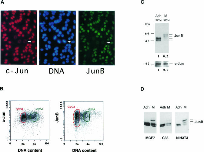
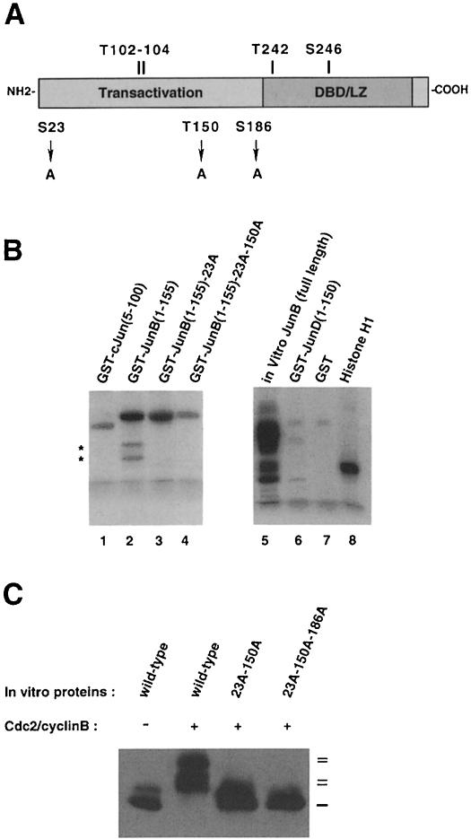
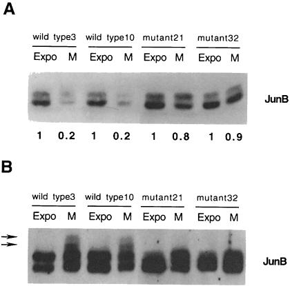
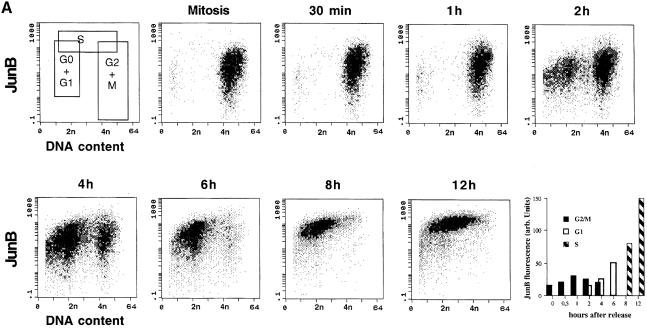
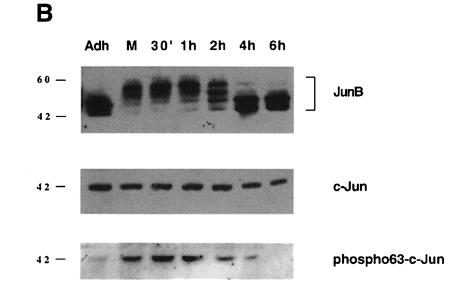
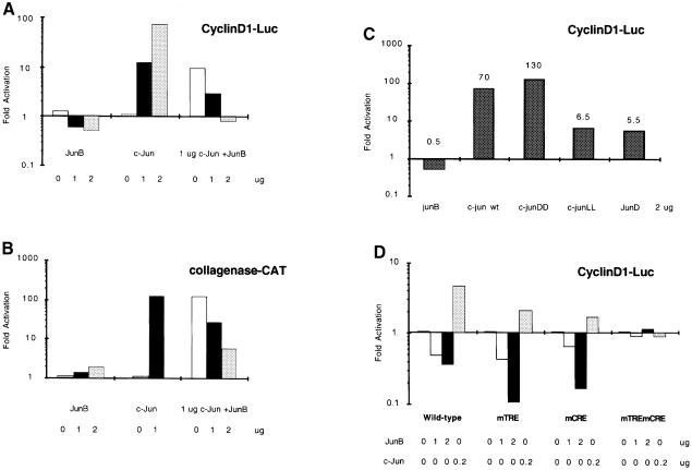
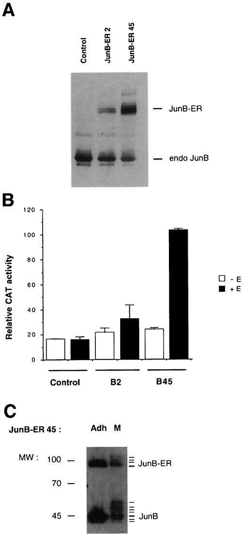
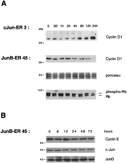
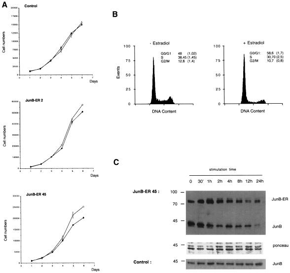
The transcription factor AP-1, composed of Jun and Fos proteins, is a major target of mitogen-activated signal transduction pathways. However, little is known about AP-1 function in normal cycling cells. Here we report that the quantity and the phosphorylation state of the c-Jun and JunB proteins vary at the M-G(1) transition. Phosphorylation of JunB by the p34(cdc2)-cyclin B kinase is associated with lower JunB protein levels in mitotic and early G(1) cells. In contrast, c-Jun levels remain constant while the protein undergoes N-terminal phosphorylation, increasing its transactivation potential. Since JunB represses and c-Jun activates the cyclin D1 promoter, these modifications of AP-1 activity during the M-G(1) transition could provide an impetus for G(1) progression by a temporal increase in cyclin D1 transcription. These findings constitute a novel example of a reciprocal connection between transcription factors and the cell cycle machinery.


Figures

None
Fig. 1. Variations of Jun protein levels during the cell cycle. (A) Co-immunofluorescence microscopy staining of exponentially growing HeLa cells for c-Jun (red), DNA (blue) and JunB (green). (B) Two-dimensional flow cytometry measurement of c-Jun and JunB levels in exponentially growing HeLa cells: FITC fluorescence (arbitrary units) of 10 000 individual cycling cells is plotted against propidium iodide fluorescence (DNA content). G0/G1 and G2/M cells are circled in green and red, respectively. (C) Analysis of c-Jun and JunB proteins in HeLa cells extracts from adherent (Adh) and nocodazole shake-off fractions (M). The percentage of 4n cells is indicated in parentheses. Equal amounts of total cell extracts were separated on SDS–PAGE and immunoprobed with the appropriate antibodies (Lallemand et al., 1997). Densitometry scan quantitation is presented below each gel. (D) Analysis of JunB proteins in different cell types. Equal amounts of extracts from adherent or M phase-enriched human breast cancer cells (MCF7), human cervical carcinoma cells (C33) and mouse fibroblasts (NIH 3T3) were probed for JunB.
None
Fig. 2. Search for JunB phosphorylating kinase. (A) Total extracts from exponentially growing, mitotic (nocodazole shake-off) and UV-treated HeLa cells were separated by SDS–PAGE and probed with antibodies specific for JunB (top), phosphorylated Jun kinase (middle) and total JNK (bottom panel). (B) c-Jun and JunB were immunoprecipitated from total cell extracts of mitotic HeLa cells and the precipitated fractions were immunoblotted for Cdc2. A non-relevant antiserum (control) was also included. (C) The Cdc2–cyclin B kinase complex induced JunB mobility shift and degradation in vitro. Starfish oocyte Cdc2–cyclin B complex was incubated for the times indicated with equal amounts of in vitro synthesized JunB protein (Iv JunB) in the presence of ATP and protease inhibitors. As a control, in vitro JunB was also incubated in the absence of Cdc2 for 240 min. The reaction mixture was resolved by SDS–PAGE and immunoprobed for JunB. The position of the different forms of JunB is shown. A cross-reacting band in the reticulocyte lysate is indicated by an asterisk.
None
Fig. 3. Mutational analysis of JunB degradation. (A) Schematic representation. Serine or threonine residues, conserved at similar positions in c-Jun, are indicated above, while serine and threonine residues specific for JunB and subject to mutational analysis are depicted below the scheme. (B) Cdc2–cyclin B phosphorylates Ser23 and Thr150 in vitro in JunB but not Thr102 and Thr104. Starfish oocyte Cdc2–cyclin B complex was incubated for 30 min with 5 µg of the indicated purified GST–Jun fusions in the presence of radioactive ATP. GST, histone H1 (0.1 µg) and reticulocyte lysate synthesized JunB were used as controls. The reactions were then resolved on SDS–PAGE and analysed with a phosphorimager. Asterisks on the left indicate degradation products. (C) Mutation of an additional residue (Ser186) in full-length JunB is necessary to abolish the Cdc2–cyclin B effect on JunB upshift. Equal amounts of in vitro sythesized JunB, JunB23A-150A and JunB23A-150A-186A were incubated with Cdc2–cyclin B. The mixture was resolved by SDS–PAGE and immunoprobed for JunB.
None
Fig. 4. (A) Mutation of Ser23, Thr150 and Thr186 stabilizes JunB in mitotic cells. Equivalent amounts of total cell extracts from exponentially growing and mitotic 293 cells stably expressing wild-type (clones 3 and 10) or mutated JunB (clones 21 and 32) were resolved by Western blot and probed for JunB. Densitometry scan quantitation is presented. (B) Mutation of Ser23, Thr150 and Thr186 reduces JunB mitotic phosphorylation. Equivalent amounts of immunoreactive JunB proteins (based on the quantification in Figure 3B) were separated by Western blot and probed for JunB. Arrows indicate the position of slower migrating bands in mitotic wild-type JunB.
None
Fig. 5. Cellular and molecular events at the G2–M–G1 transition. Synchronized populations of HeLa cells were analysed by two-dimensional flow cytometry and Western blot. (A) Flow cytometry: variation of JunB during cell cycle progression of nocodazole-released cells. JunB-specific fluorescence is plotted against DNA content or (last panel) against time. The first panel (top left) indicates the limits of each subpopulation of cells. The positions of 2n and 4n cells are indicated. The last panel is based on JunB fluorescence peak values, obtained after gating the different populations. (B) Western blot analysis of JunB (the film was overexposed to reveal the slower migrating JunB bands), c-Jun and Ser63 phosphorylated c-Jun in cell extracts corresponding to the same time points.
None
Fig. 6. Regulation of the human cyclin D1 promoter by Jun proteins. (A) JunB counteracts c-Jun activation of the cyclin D1 gene promoter. HeLa cells were transiently transfected with the Δ973cyclinD1LUC plasmid together with increasing amounts of expression constructs for c-Jun or JunB and a combination of 1 µg of c-Jun and increasing amounts of JunB. (B) JunB is a weak but positive transactivator on a canonical TRE-responsive promoter. In the same type of experiment, HeLa cells were transfected with a collagenase–CAT construct together with expression constructs for c-Jun and JunB. (C) Effect of each of the Jun proteins on the cyclin D1 gene promoter. HeLa cells were transiently transfected with the Δ973cyclinD1LUC plasmid together with expression constructs for c-Jun, JunB, JunD and mutants of c-Jun in the N-terminal phosphorylation sites. (D) JunB and c-Jun affect the same sites on the cyclin D1 promoter. HeLa cells were transiently transfected with Δ973cyclinD1LUC, Δ973cyclinD1mTRELUC or Δ973cyclinD1mCRELUC plasmids together with c-Jun and JunB expression vectors.The mean results of two representative experiments are presented.
None
Fig. 7. (A) Immunoblotting analysis of JunB proteins in stable JunB–ER clones. Equal amounts of total extracts from β-oestradiol-stimulated NIH 3T3 control cells, NIH 3T3/JunB–ER clone 2 and clone 45 were separated by SDS–PAGE and probed for JunB. Labelled lanes indicate the position of the endogenous JunB protein and the ectopically expressed JunB–ER fusion protein of ∼80 kDa. (B) The JunB–ER protein activates transcription of a transiently transfected TRE reporter in a hormone-dependent fashion. Control and JunB–ER cells (clones B2 and B45) were transiently transfected with a collagenase–CAT construct and treated for 40 h with β-oestradiol or solvent. The values and standard deviation represent the results of three independent experiments. (C) The JunB–ER fusion protein is also subjected to Cdc2–cyclin B phosphorylation during mitosis. Equal amounts of total extracts from adherent (Adh) or mitotic (M) NIH 3T3/JunB–ER45 cells were separated by SDS–PAGE and probed for JunB.
None
Fig. 8. Western blotting analysis of NIH 3T3/JunB–ER-45 and NIH 3T3/c-Jun-ER cells. (A) Equivalent amounts of total cell extracts from stimulated cells were separated by SDS–PAGE, transferred to nitrocellulose membranes and probed with a mouse monoclonal anti-cyclin D1 antibody. In the case of JunB–ER, the filter was Ponceau stained to check the correct protein loading and reprobed with a monoclonal anti-Rb. (B) JunB–ER induction does not affect either cyclin E, c-Jun or JunD expression. Equivalent amounts of total cell extracts were subjected similarly to Western blot with the appropriate antibodies.
None
Fig. 9. (A) Effect of the JunB–ER protein on the growth of NIH 3T3 cells. Control NIH 3T3 and NIH 3T3/JunB–ER clones were cultured in the presence (closed circles) or absence (open circles) of 1 µM β-oestradiol. Cells were counted daily and the mean values of triplicate cultures have been plotted against time. (B) Cell cycle distribution of 48 h untreated and β-oestradiol-treated NIH 3T3/JunB–ER cells (clone B45). The DNA content is presented as relative fluorescence, and a quantitation of the results is presented. (C) Immunoblotting analysis of the JunB proteins during β-oestradiol stimulation of NIH 3T3/JunB–ER-45 or NIH 3T3 control cells. Equal amounts (documented for JunB–ER by Ponceau staining) of nuclear extracts from cells stimulated with β-oestradiol were probed for JunB. The endogenous JunB and the ectopically expressed JunB–ER fusion protein are indicated.

References

    1. Albanese C., Johnson,J., Watanabe,G., Eklund,N., Vu,D., Arnold,A. and Pestell,R.G. (1995) Transforming p21ras mutants and c-Ets-2 activate the cyclin D1 promoter through distinguishable regions. J. Biol. Chem., 270, 23589–23597. - PubMed
    1. Beier F., Lee,R.J., Taylor,A.C., Pestell,R.G. and LuValle,P. (1999) Identification of the cyclin D1 gene as a target of activating transcription factor 2 in chondrocytes. Proc. Natl Acad. Sci. USA, 96, 1433–1438. - PMC - PubMed
    1. Binetruy B., Smeal,T. and Karin,M. (1991) Ha-Ras augments c-Jun activity and stimulates phosphorylation of its activation domain. Nature, 351, 122–127. - PubMed
    1. Bossy-Wetzel E., Bakiri,L. and Yaniv,M. (1997) Induction of apoptosis by the transcription factor c-Jun. EMBO J., 16, 1695–1709. - PMC - PubMed
    1. Boulikas T. (1995) Phosphorylation of transcription factors and control of the cell cycle. Crit. Rev. Eukaryot. Gene Expr., 5, 1–77. - PubMed

MeSH terms