. 2000 May 2;19(9):2115-26.
doi: 10.1093/emboj/19.9.2115.

Rcl1p, the yeast protein similar to the RNA 3'-phosphate cyclase, associates with U3 snoRNP and is required for 18S rRNA biogenesis

Affiliations

Rcl1p, the yeast protein similar to the RNA 3'-phosphate cyclase, associates with U3 snoRNP and is required for 18S rRNA biogenesis

E Billy et al. EMBO J. .

Abstract

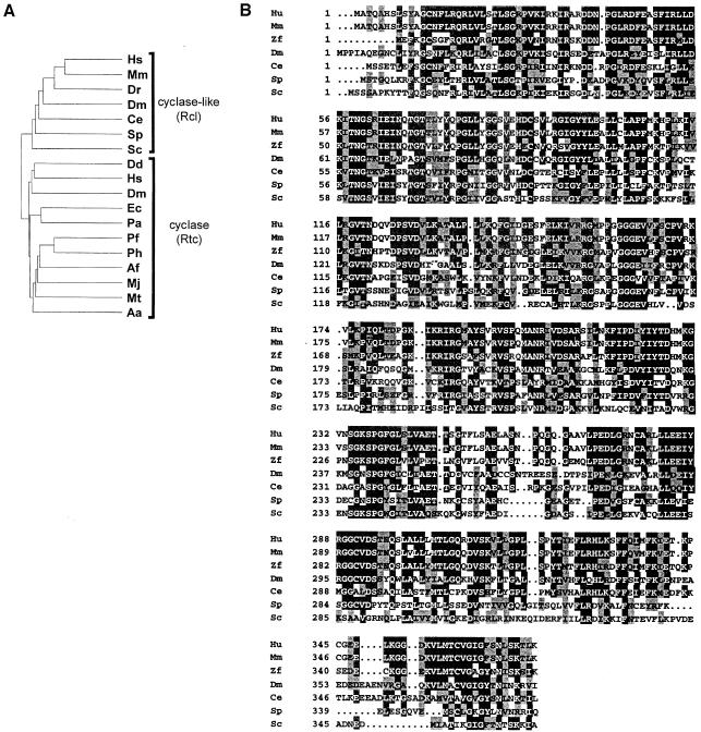
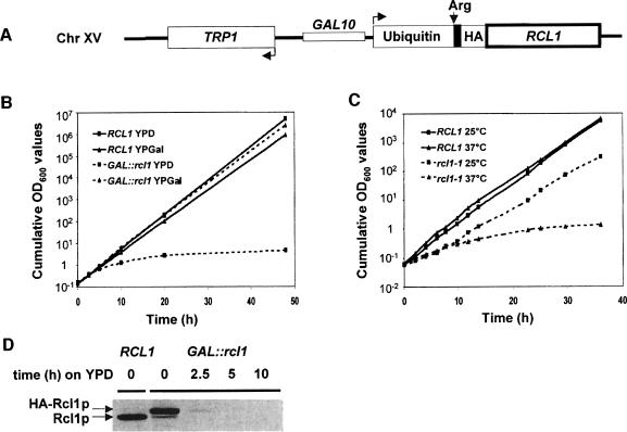
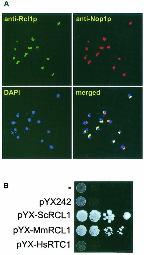
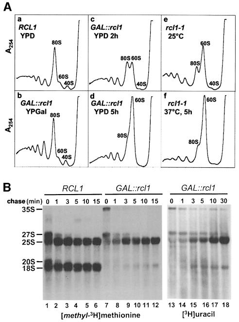
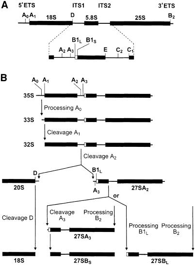
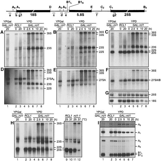
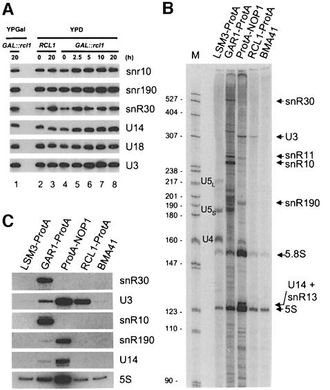
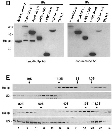
RNA 3'-terminal phosphate cyclases are evolutionarily conserved enzymes catalysing conversion of the 3'-terminal phosphate in RNA to the 2',3'-cyclic phosphodiester. Their biological role remains unknown. The yeast Saccharomyces cerevisiae contains a gene encoding a protein with strong sequence similarity to the characterized cyclases from humans and Escherichia coli. The gene, named RCL1 (for RNA terminal phosphate cyclase like), is essential for growth, and its product, Rcl1p, is localized in the nucleolus. Depletion or inactivation of Rcl1p impairs pre-rRNA processing at sites A(0), A(1) and A(2), and leads to a strong decrease in 18S rRNA and 40S ribosomal subunit levels. Immunoprecipitations indicate that Rcl1p is specifically associated with the U3 snoRNP, although, based on gradient analyses, it is not its structural component. Most of Rcl1p sediments in association with the 70-80S pre-ribosomal particle and a 10S complex of unknown identity. Proteins similar to Rcl1p are encoded in genomes of all eukaryotes investigated and the mouse orthologue complements yeast strains depleted of Rcl1p. Possible functions of Rcl1p in pre-rRNA processing and its relationship to the RNA 3'-phosphate cyclase are discussed.


Figures

None
Fig. 1. Dendritic tree of cyclases (Rtc) and cyclase-like proteins (Rcl) from different organisms (A) and comparison of amino acid sequences of proteins belonging to the Rcl subfamily (B). The compared Rtc proteins originate from the following organisms: Hs, Homo sapiens [SwissProt (SP) O00442]; Dm, Drosophila melanogaster (GenBank SPTREMBL O77264); Dd, Dictyostelium discoideum (SP AAB70847); Ec, E.coli (SP P46849); Pa, Pseudomonas aeruginosa (The Pseudomonas Genome Project); Ph, Pyrococcus horikoshi (Entrez BAA30639); Pf, Pyrococcus furiosus (The Insitute for Genomic Research, Gaithersburg, MD); Af, Archaeoglobus fulgidus (AAB89810); Mj, Methanococcus jannaschii (SP Q60335); Mt, Methanobacterium thermoautotrophicum (AAB86375); Aa, Aquifex aeolicus (AAC06852). Compared Rcl proteins originate from: Hs, H.sapiens (Genschik et al., 1997; GenBank AJ276894); Mm, Mus musculus (GenBank AJ276895); Dr, Danio rerio (zebrafish; GenBank ESTs: AW078116.1 and AW059073.1); Dm, D.melanogaster (SP P56175); Ce, Caenorhabditis elegans (SP Q23400); Sp, Schizosaccharomyces pombe (SP Q09870); Sc, S.cerevisiae (SP Q08096). Multiple sequence alignment was performed with the ClustalW program as described (Genschik et al., 1997). Identical amino acids and amino acids conserved in at least 50% of sequences are indicated by black and grey boxes, respectively.
None
Fig. 2. Conditional alleles. (A) Scheme of the GAL::rcl1 allele. The RCL1 ORF is fused to the ubiquitin, HA-tag and LacI (shown in black) regions. Following removal of ubiquitin, the fusion protein starts with the destabilizing amino acid, arginine (Arg). (B) Growth curves of GAL::rcl1 and the wild-type strain in YPD and YPGal. Cells were shifted to non-permissive conditions at time 0. (C) Growth curves of rcl1-1 ts and wild-type strains at 25 and 37°C. (D) Depletion of HA-Rcl1p analysed by Western blotting with α-Rcl1p Abs. Positions of the HA-Rcl1p and wild-type Rcl1p proteins are indicated.
None
Fig. 3. Nucleolar localization of Rcl1p and complementation of the yeast GAL::rcl1 mutant by the mouse Rcl1p-like protein. (A) Rcl1p in the BMA41 strain was detected by a rabbit α-Rcl1p Ab, followed by a goat FITC-coupled α-rabbit Ab. Nop1p was detected by mouse mAb A66 followed by a goat TR-conjugated α-mouse Ab. Chromatin was stained with DAPI. Merging of all three images is shown in the bottom right panel. (B) The GAL::rcl1 strain was transformed with either pYX-ScRCL1, pYX-MmRCL1, pYX-HsRTC1 or the empty vector pYX242; upper row, untransformed cells. Complementation was assayed by monitoring growth on YPD plates. Western blotting with Abs against the human cyclase verified that it is expressed in cells transformed with pYX-HsRTC1 (data not shown).
None
Fig. 4. Polysome profile (A) and pulse–chase (B) analyses. (A) Polysome profiles of GAL::rcl1 (panels b–d), rcl1-1 ts (panels e–f) mutant strains and a wild-type (panel a) strain grown under permissive and/or restrictive conditions. Time of growth on YPD or at the restrictive temperature is indicated. Polysome profiles of wild-type cells grown at 30°C (panel a), and at 25 and 37°C (not shown) are similar. (B) Pulse–chase labelling of rRNA with [methyl-3H]methionine (lanes 1–12) and [3H]uracil (lanes 13–18) in the wild-type and GAL::rcl1 strains. Labelling (3 min) was performed following 5 h growth on YPD.
None
Fig. 5. Pre-rRNA processing pathway in yeast. (A) Structure of 35S pre-rRNA. (B) Physiological pathway of 35S pre-rRNA processing. Final steps of 5.8 and 25S maturation are not shown. Processing sites and intermediates are indicated. For more details, see Venema and Tollervey (1995); Kressler et al. (1999).
None
Fig. 6. Depletion or inactivation of Rcl1p affects pre-rRNA processing at sites A0, A1 and A2 (A–G and H, lanes 1–8). Northern analysis of pre-rRNAs accumulating in the wild-type strain grown in YPD (lanes 2 and 3) and GAL::rcl1 strain grown either in YPGal (lanes 1) or YPD (lanes 4–8). The following oligonucleotide probes (indicated schematically above the gels) were used: (A) probe a, the 5′ETS upstream of A0; (B) probe b, junction of 5′ETS and 18S rRNA; (C and H) probe c, the ITS1 upstream of A2; (D) probe d, between A2 and A3; (E) probe e, the ITS1 downstream of A3; (F) probe f, the ITS2 upstream of C2; (G) probes g1 and g2, complementary to 18S and 25S rRNA, respectively; (H) lanes 9–12, processing intermediates accumulating in the rcl1-1 ts mutant grown at permissive or restrictive temperature (37°C, 5 h). (I) Primer extension analysis of RNAs accumulating in the GAL::rcl1 strain grown either in YPGal (lane 1) or YPD (lanes 3–7), and in the wild-type strain grown in YPD (lane 2). Oligo g1 was used for primer extension to sites A0 and A1, and oligo f for extension to sites A2, A3 and B1L/S. Positions of primer extension stops corresponding to different pre-rRNA cleavage sites are indicated. The A0 and A1 panels represent different exposures of the same gel.
None
Fig. 7. Rcl1p is specifically associated with but is not a structural component of U3 snoRNP. (A) Depletion of Rcl1p has no effect on snoRNA levels. For lane description, see Figure 6. (B and C) Rcl1p immunoprecipitates U3 snoRNA. In (B) RNA isolated from IP pellets was labelled with [5′-32P]pCp and fractionated by 6% PAGE. U6 snRNA immunoprecipitated by Lsm3p is not labelled with [5′-32P]pCp (Lund and Dahlberg, 1992). In (C) the same material was separated by 8% PAGE and analysed by Northern blotting. The amount of U3 precipitated by Rcl1p-ProtA is three times less than that precipitated by ProtA-Nop1p. This is consistent with the observations that only ∼50% of U3 RNA is associated with large complexes co-sedimenting with Rcl1p (see E) and that the efficiency of Rcl1p-ProtA binding to IgG–Sepharose is lower than that of other ProtA fusions, including ProtA-Nop1p (data not shown). (D) Co-immunoprecipitation of U3 snoRNP proteins and Rcl1p. Eluates from IPs carried out with the strains indicated were analysed by Western blotting using α-Rcl1p Ab (left panel) and non-immune Ab detecting only ProtA-tagged proteins (right panel). (E) Sedimentation of Rcl1p and U3snoRNP on 10–30% glycerol gradients run for long (upper panel) and short (lower panel) times. Positions of 80S, 60S and 40S ribosomes and protein markers (from Pharmacia Biotech) run in parallel gradients (19S thyroglobulin, 669 kDa; 11.3S catalase, 232 kDa; 8S aldolase, 158 kDa; 4.3S bovine serum albumin, 67 kDa) are indicated. Apparently faster sedimentation of the 10S Rcl1p peak upon shorter centrifugation (lower panel) may be due to either poor resolution of different Rcl1p-containing complexes or retention of loosely associated protein(s) that is lost during prolonged centrifugation. We have confirmed that the 43S peak corresponds to the precursor of the 40S subunit by demonstrating that it co-fractionates with 20S precursor rRNA; its association with U3 RNA suggests that it represents a nuclear form of the 43S particle (Trapman et al., 1975).

Similar articles

Cited by

References

    1. Arn E.A. and Abelson,J.N. (1998) RNA ligases: function, mechanism and sequence conservation. In Simons,R.W. and Grunberg-Manago,M. (eds), RNA Structure and Function. Cold Spring Harbor Laboratory Press, Cold Spring Harbor, NY, pp. 695–726.
    1. Balakin A.G., Smith,L. and Fournier,M.J. (1996) The RNA world of the nucleolus: two major families of small RNAs defined by different box elements with related functions. Cell, 86, 823–834. - PubMed
    1. Baudin A., Ozier-Kalogeropoulos,O., Denouel,A., Lacroute,F. and Cullin,C. (1993) A simple and efficient method for direct gene deletion in S.cerevisiae. Nucleic Acids Res., 21, 3329–3330. - PMC - PubMed
    1. Beltrame M. and Tollervey,D. (1992) Identification and functional analysis of two U3 binding sites on yeast pre-ribosomal RNA. EMBO J., 11, 1531–1542. - PMC - PubMed
    1. Beltrame M. and Tollervey,D. (1995) Base pairing between U3 and the pre-ribosomal RNA is required for 18S rRNA synthesis. EMBO J., 14, 4350–4356. - PMC - PubMed

MeSH terms

Associated data