. 2000 May 1;191(9):1467-76.
doi: 10.1084/jem.191.9.1467.

The neutrophil-activating protein (HP-NAP) of Helicobacter pylori is a protective antigen and a major virulence factor

Affiliations

The neutrophil-activating protein (HP-NAP) of Helicobacter pylori is a protective antigen and a major virulence factor

B Satin et al. J Exp Med. .

Abstract

Helicobacter pylori infection induces the appearance of inflammatory infiltrates, consisting mainly of neutrophils and monocytes, in the human gastric mucosa. A bacterial protein with neutrophil activating activity (HP-NAP) has been previously identified, but its role in infection and immune response is still largely unknown. Here, we show that vaccination of mice with HP-NAP induces protection against H. pylori challenge, and that the majority of infected patients produce antibodies specific for HP-NAP, suggesting an important role of this factor in immunity. We also show that HP-NAP is chemotactic for human leukocytes and that it activates their NADPH oxidase to produce reactive oxygen intermediates, as demonstrated by the translocation of its cytosolic subunits to the plasma membrane, and by the lack of activity on chronic granulomatous disease leukocytes. This stimulating effect is strongly potentiated by tumor necrosis factor alpha and interferon gamma and is mediated by a rapid increase of the cytosolic calcium concentration. The activation of leukocytes induced by HP-NAP is completely inhibited by pertussis toxin, wortmannin, and PP1. On the basis of these results, we conclude that HP-NAP is a virulence factor important for the H. pylori pathogenic effects at the site of infection and a candidate antigen for vaccine development.


Figures

Figure 1
Figure 1
HP-NAP is immunogenic in humans and protective in mice. (A) The sera of an H. pylori–infected patient 1 and a negative control 2 were assayed by immunoblotting followed by development with enhanced chemiluminescence on samples of H. pylori total sonicates (left lanes) and purified recombinant HP-NAP (right lanes), subjected to SDS-PAGE, and then transferred onto nitrocellulose paper. Numbers on the left refers to the position of molecular weight markers (in kD). The last lane (αNAP) shows an immunoblot of recombinant HP-NAP stained with an anti–HP-NAP antiserum obtained from rabbits. (B) Groups of 10 CD-1 mice were immunized three times intragastrically with the indicated antigens together with the LTK63 mutant as a mucosal adjuvant, and then challenged with 109 CFU of H. pylori strain SPM326 (see Materials and Methods for details). Mice were considered protected when no colonies were counted from their stomachs.
Figure 3
Figure 3
HP-NAP upregulates the expression of β2 integrins in neutrophils and monocytes. The HP-NAP–induced expression of the protein on the cell surface was determined by FACS® analysis with a specific mAb: (A) control cells; (B) cells treated with 0.2 mM FMLP for 30 min at 37°C; (C) cells treated with 0.5 μM HP-NAP for 30 min at 37°C.
Figure 4
Figure 4
Stimulation of H2O2 production by different doses of HP-NAP in human peripheral blood neutrophils (A) and monocytes (B). H2O2 was measured with the homovanillic acid method (reference 34). Very similar results were obtained by assaying the reduction of cytochrome c (not shown). Points are the average of five different experiments run in triplicate, and bars represent SD values. Symbols indicate: ▪, control; ♦, the data obtained with HP-NAP purified from H. pylori (average value of data obtained from two different preparations); all other points shown here and in the following pictures were obtained with highly purified recombinant HP-NAP expressed in B. subtilis: ▴, 0.6 μM HP-NAP; ▾, 1.2 μM HP-NAP; •, 3 μM HP-NAP; ○, 1 μM FMLP.
Figure 5
Figure 5
IFN-γ (A) and TNF-α (B) potentiate the stimulation of ROI production by HP-NAP. H2O2 was measured with the homovanillic acid method (reference 34). Before addition of 1.2 μM HP-NAP, neutrophils were preincubated for 60 min with 5 ng/ml IFN-γ or 15 min with 5 ng/ml TNF-α at 37°C. Points are the average of three different experiments run in triplicate, and bars represent SD values.
Figure 6
Figure 6
Translocation of cytosolic components of NADPH oxidase on the plasma membrane in human neutrophils stimulated with HP-NAP. Lane 1, unstimulated neutrophils; lane 2, neutrophils stimulated for 15 min with 3 μM HP-NAP; lane 3, neutrophils stimulated with PMA (50 ng/ml) for 15 min. The conditions of cell stimulation, fractionation, and detection of cells components are described in Materials and Methods.
Figure 7
Figure 7
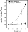
HP-NAP does not stimulate H2O2 production in CGD neutrophils defective of NADPH oxidase. Experiments were performed twice in triplicate, and H2O2 production was measured with the homovanillic acid method (reference 34). Bars represent the SD value.
Figure 8
Figure 8
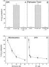
Effect of DPI (A), pertussis toxin (B), wortmannin (C), and PP1 (D) on the stimulation of H2O2 production by HP-NAP. Human neutrophils were preincubated with the indicated inhibitor at the concentrations reported in the figure for 5 min at 37°C (2 h for pertussis toxin), before addition of 3 μM HP-NAP, and then assayed as in the legend to Fig. 5. Data are the average of the results of two different experiments run in triplicate, and bars represent SD values.
Figure 9
Figure 9
Effect of HP-NAP on [Ca2+]i in neutrophils in the absence or presence of pertussis toxin, wortmannin, PP1, and EGTA. (A) Human neutrophils loaded with Fura-2 for 25 min were washed and then exposed to the given concentrations of HP-NAP in a stirred fluorimeter cuvette. Fluorescence outputs were converted to [Ca2+] after calibration. (B) Only part of the increase in cytosolic [Ca2+] triggered by HP-NAP (solid line) is due to release from intracellular stores (broken line). (C) Pertussis toxin completely prevents the HP-NAP–induced rise in cytosolic [Ca2+], whereas other inhibitors are ineffective.
Figure 2
Figure 2
Dose–response stimulation of HP-NAP chemotaxis in neutrophils and monocytes. The chemotactic activity of HP-NAP on human neutrophils and monocytes is compared with that of FMLP. Chemotaxis was measured in Costar transwells as specified in the Materials and Methods. Bars represent the average of three independent experiments performed in duplicates, and the SD values are given at the top.

Comment in

References

    1. Warren J.D., Marshall B.J. Unidentified curved bacilli on gastric epithelium in active chronic gastritis. Lancet. 1983;1:1273–1275. - PubMed
    1. Marshall B.J., Armstrong J.A., McGechie D.B., Glancy R.J. Attempt to fulfill Koch's postulates for pyloric Campylobacter . Med. J. Austr. 1985;142:436–439. - PubMed
    1. Goodwin C.S. Helicobacter pylori gastritis, peptic ulcer and gastric cancerclinical and molecular aspects. Clin. Infect. Dis. 1997;25:1017–1019. - PubMed
    1. Bayerdorffer E., Lehn N., Hatz R., Mannes G.A., Oertel H., Sauerbruch T., Stolte M. Difference in expression of Helicobacter pylori gastritis in antrum and body. Gastroenterology. 1992;102:1575–1582. - PubMed
    1. Fiocca R., Villani L., Luinetti O., Gianotti A., Perego M., Alvisi C., Turpini F., Solcia E. Helicobacter colonization and histopathological profile of chronic gastritis in patients with or without dyspepsia, mucosal erosion and peptic ulcera morphological approach to the study of ulcerogenesis in man. Virchows Archiv. A Pathol. Anat. Histopathol. 1992;420:489–492. - PubMed

Publication types

MeSH terms