. 2000 May 1;149(3):537-46.
doi: 10.1083/jcb.149.3.537.

Epsin 1 undergoes nucleocytosolic shuttling and its eps15 interactor NH(2)-terminal homology (ENTH) domain, structurally similar to Armadillo and HEAT repeats, interacts with the transcription factor promyelocytic leukemia Zn(2)+ finger protein (PLZF)

Affiliations

Epsin 1 undergoes nucleocytosolic shuttling and its eps15 interactor NH(2)-terminal homology (ENTH) domain, structurally similar to Armadillo and HEAT repeats, interacts with the transcription factor promyelocytic leukemia Zn(2)+ finger protein (PLZF)

J Hyman et al. J Cell Biol. .

Abstract

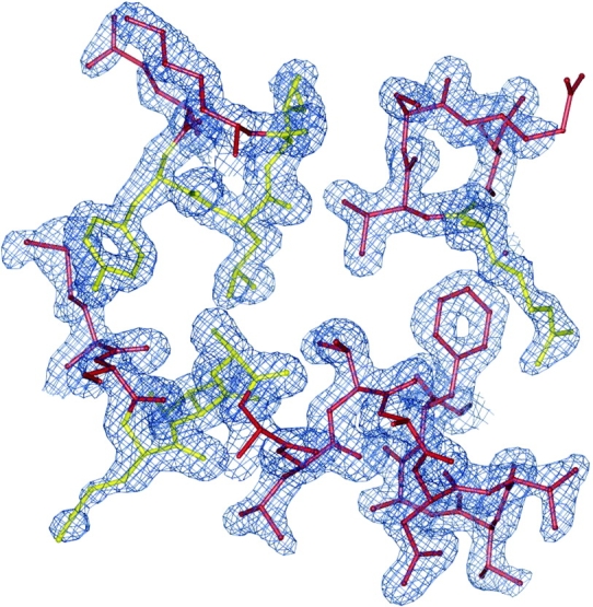
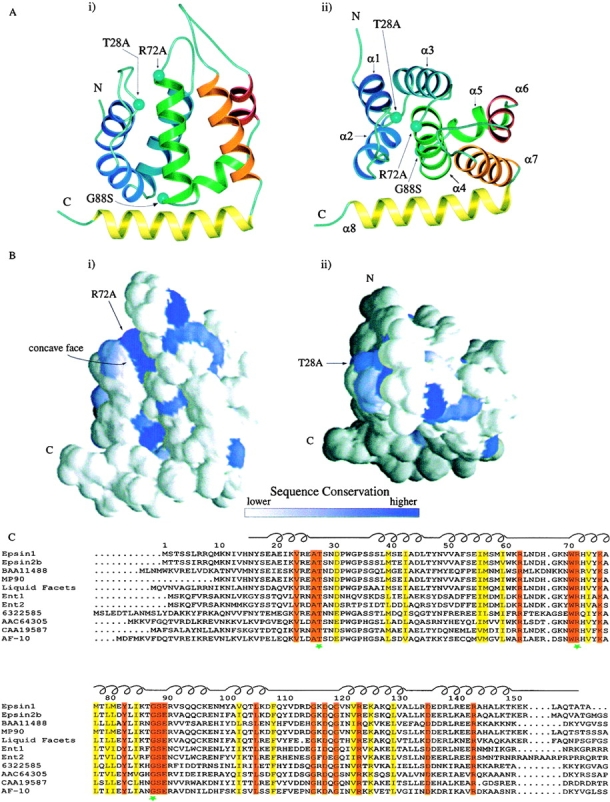
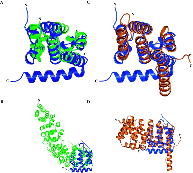
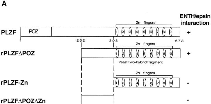
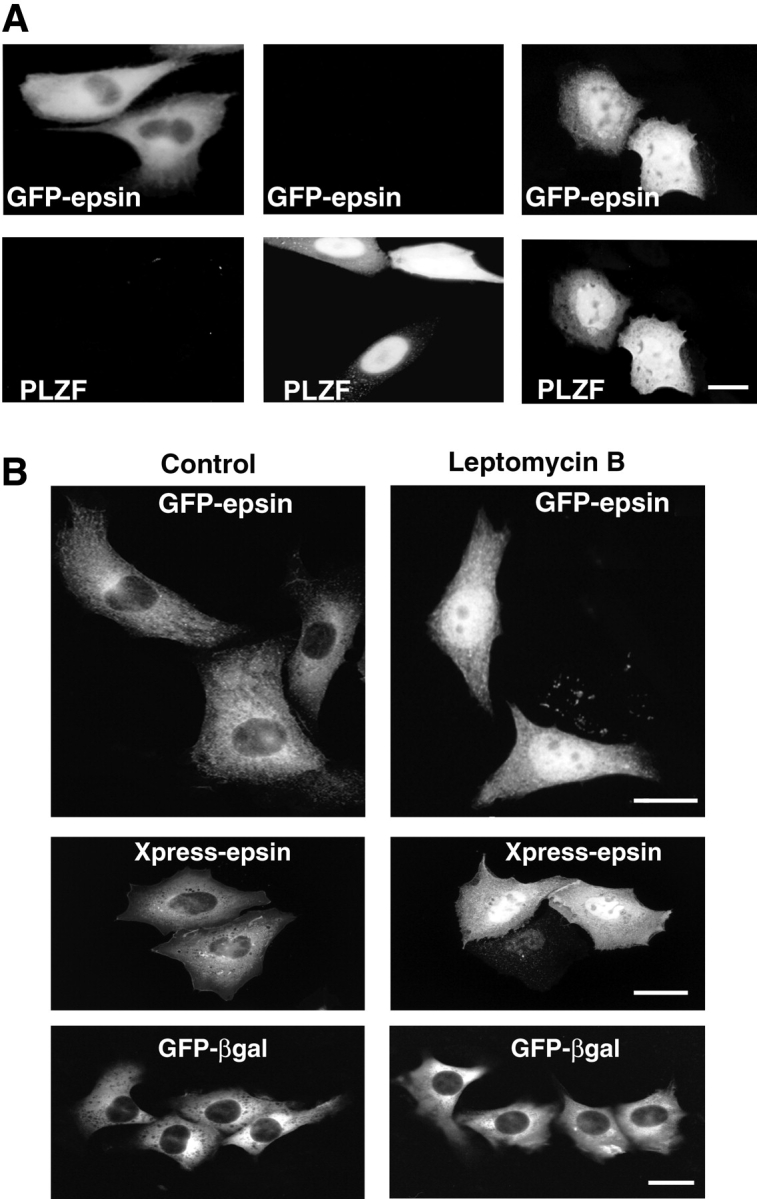
Epsin (Eps15 interactor) is a cytosolic protein involved in clathrin-mediated endocytosis via its direct interactions with clathrin, the clathrin adaptor AP-2, and Eps15. The NH(2)-terminal portion of epsin contains a phylogenetically conserved module of unknown function, known as the ENTH domain (epsin NH(2)-terminal homology domain). We have now solved the crystal structure of rat epsin 1 ENTH domain to 1.8 A resolution. This domain is structurally similar to armadillo and Heat repeats of beta-catenin and karyopherin-beta, respectively. We have also identified and characterized the interaction of epsin 1, via the ENTH domain, with the transcription factor promyelocytic leukemia Zn(2)+ finger protein (PLZF). Leptomycin B, an antifungal antibiotic, which inhibits the Crm1- dependent nuclear export pathway, induces an accumulation of epsin 1 in the nucleus. These findings suggest that epsin 1 may function in a signaling pathway connecting the endocytic machinery to the regulation of nuclear function.


Figures

Figure 1
Figure 1
Representative experimental electron density map for residues 30–44, 72–76, and 81–85 contoured at 1.4 Å obtained by MAD phasing and subsequent density modification. The final refined model is shown using a ball and stick representation, with residues in yellow indicating a high degree of sequence conservation. The backbone trace was clearly visible for a portion of α helix 2, and exceptional density was found for most side chains. Figure generated using MOLSCRIPT (Kraulis 1991).
Figure 2
Figure 2
Overall structure of the ENTH domain and sequence alignment. A, Ribbon drawings of the ENTH domain. The drawing at right (ii) is related to the drawing at left (i) by a 60° rotation towards the viewer. The eight α helices of the ENTH domain are indicated by different colors. The T28A, R72A, and G88S mutations made in this study are indicated by aquamarine balls. α Helices 1–8 are indicated in i. Figures generated using MOLSCRIPT (Kraulis 1991). B, Sequence conservation projected onto accessible surface representations of the ENTH domain. These surface representations correspond to the same two views (i and ii) in A. Figures generated using GRASP (Nicholls, 1991). C, Sequence alignment of ENTH domains from various species. Identical residues are indicated in red, conserved residues in yellow. Green stars indicate residues that have been mutated in this study. The schematic diagram above the alignment indicates the location of loop and helix regions. Accession number and species as follows: epsin 1, AAC33823, Rattus norvegicus; epsin 2b, AAC78609, Homo sapiens; BAA11488, Homo sapiens; MP90, AAC60123, Xenopus laevis; liquid facets, AAF05113, Drosophila melanogaster; Ent1, 6320039, S. cerevisiae; Ent2, 6323235, S. cerevisiae; 6322585, S. cerevisiae; AAC64305, Arabidopsis thaliana; CAA19587, Schizosaccharomyces pombe; AF-10, AAB68030, Avena fatua. Alignment generated using CLUSTAL W (Thompson et al. 1994) and AMAS (Livingston and Barton 1993).
Figure 3
Figure 3
Alignment of structurally similar protein folds. A, Alignment of the ENTH domain of epsin 1 (blue) with one portion of the armadillo repeat region of β-catenin (green), showing a good overall superimposition between α helices in the central region of the domain. This alignment shows the ENTH domain in the same orientation as in Fig. 2A ii. B, Same as A, but showing the entire β-catenin armadillo region and the ENTH domain in the same orientation as in Fig. 2A i. C, Alignment of the ENTH domain (blue) with a segment of karyopherin-β (orange). This alignment shows the ENTH domain in the same orientation as in Fig. 2A ii. D, Same as C, showing the entire structure of karyopherin-β and the ENTH domain in the orientation of Fig. 2A i. Structural alignments produced using WHATIF (Vriend 1990). Figures generated using MOLSCRIPT (Kraulis et al., 1991).
Figure 4
Figure 4
Interaction between the ENTH domain of epsin 1 and PLZF. A, Schematic representation of human PLZF and of the rat PLZF fragments discussed in this study. Numbers indicate amino acid numbers. B, Interaction of the GST-ENTH domain fusion protein with rPLZFΔPOZ. An extract of CHO cells transfected with flag-tagged rPLZFΔPOZ was affinity-purified on either GST or a GST-ENTH domain fusion protein. The starting material and the bead-bound material were analyzed by Western blotting using antibody directed against the flag epitope. C, ENTH domain affinity purification from a rat brain extract. A Triton X-100 extract of a rat brain total homogenate was affinity-purified on ENTH domain conjugated to beads or control beads. The starting material and the bead-bound material were analyzed by Western blotting for PLZF. D and E, PLZF and epsin can be coprecipitated from a rat brain Triton X-100 extract. D, Immuno-precipitates generated by antiepsin 1 antibodies or control IgGs were analyzed by Western blotting with anti-rat PLZF or anti-p53 (used as control) antibodies. E, Immunoprecipitates generated by anti-PLZF antibodies or control IgGs were analyzed by Western blotting using antiepsin 1 antibodies. F: Mutant ENTH domains fail to interact with PLZF. An extract of CHO cells transfected with flag-tagged human PLZF was affinity-purified on wild-type or mutant (T28A, R72A, and G88S) GST-ENTH domain fusion proteins. The starting material, the bead-unbound and the bead-bound material were analyzed by Western blotting using antibody directed against the flag epitope or anticlathrin antibody as a control. B, Bead-bound; c, control IgGs; e, antiepsin antibodies; P, pellet; S, supernatant; SM, starting material; U, bead-unbound.
Figure 4
Figure 4
Interaction between the ENTH domain of epsin 1 and PLZF. A, Schematic representation of human PLZF and of the rat PLZF fragments discussed in this study. Numbers indicate amino acid numbers. B, Interaction of the GST-ENTH domain fusion protein with rPLZFΔPOZ. An extract of CHO cells transfected with flag-tagged rPLZFΔPOZ was affinity-purified on either GST or a GST-ENTH domain fusion protein. The starting material and the bead-bound material were analyzed by Western blotting using antibody directed against the flag epitope. C, ENTH domain affinity purification from a rat brain extract. A Triton X-100 extract of a rat brain total homogenate was affinity-purified on ENTH domain conjugated to beads or control beads. The starting material and the bead-bound material were analyzed by Western blotting for PLZF. D and E, PLZF and epsin can be coprecipitated from a rat brain Triton X-100 extract. D, Immuno-precipitates generated by antiepsin 1 antibodies or control IgGs were analyzed by Western blotting with anti-rat PLZF or anti-p53 (used as control) antibodies. E, Immunoprecipitates generated by anti-PLZF antibodies or control IgGs were analyzed by Western blotting using antiepsin 1 antibodies. F: Mutant ENTH domains fail to interact with PLZF. An extract of CHO cells transfected with flag-tagged human PLZF was affinity-purified on wild-type or mutant (T28A, R72A, and G88S) GST-ENTH domain fusion proteins. The starting material, the bead-unbound and the bead-bound material were analyzed by Western blotting using antibody directed against the flag epitope or anticlathrin antibody as a control. B, Bead-bound; c, control IgGs; e, antiepsin antibodies; P, pellet; S, supernatant; SM, starting material; U, bead-unbound.
Figure 5
Figure 5
Immunofluorescence micrographs demonstrating that epsin may accumulate in the nucleus. A, Overexpression of human PLZF induces a partial accumulation of epsin in the nucleus. CHO cells were transiently transfected with epsin alone, human PLZF alone, or both proteins together, and then analyzed by confocal microscopy immunofluorescence. A pool of epsin is localized in the nucleus only in cells expressing both proteins. B, Leptomycin B induces an accumulation of epsin in the nucleus. CHO cells were transiently transfected with GFP-epsin, Xpress-tagged epsin, or the control protein GFP-β-galactosidase (GFP-βgal) and incubated for 3.5 h in the presence of leptomycin B before microscopic analysis. An accumulation of epsin is observed in the nucleus only in cells treated with leptomycin B. GFP-β-galactosidase was only cytosolic both in the presence and in the absence of leptomycin B. Bars, 12 μm.

References

    1. Abrahams J.P., Leslie A.G.W. Methods used in the structure determination of bovine mitochondrial F1 ATPase. Acta Crystallogr. 1996;D52:30–42. - PubMed
    1. Barth A.I.M., Näthke I.S., Nelson W.J. Cadherins, catenins and APC proteininterplay between cytoskeletal complexes and signalling pathways. Curr. Opin. Cell Biol. 1997;9:683–690. - PubMed
    1. Benmerah A., Lamaze C., Bègue B., Schmid S.L., Dautry-Varsat A., Cerf-Bensussan N. AP-2/Eps15 interaction is required for receptor mediated endocytosis. J. Cell Biol. 1998;140:1055–1062. - PMC - PubMed
    1. Bricogne G. Bayesian statistical viewpoint on structure determinationbasic concepts and examples. Methods Enzymol. 1997;276:361–423. - PubMed
    1. Brunger A.T. Free R valuea novel statistical quantity for assessing the accuracy of crystal structures. Nature. 1992;355:472–475. - PubMed

Publication types

MeSH terms

Associated data