. 2000 May 1;149(3):647-56.
doi: 10.1083/jcb.149.3.647.

Drosophila beta spectrin functions independently of alpha spectrin to polarize the Na,K ATPase in epithelial cells

Affiliations

Drosophila beta spectrin functions independently of alpha spectrin to polarize the Na,K ATPase in epithelial cells

R R Dubreuil et al. J Cell Biol. .

Abstract

Spectrin has been proposed to function as a sorting machine that concentrates interacting proteins such as the Na,K ATPase within specialized plasma membrane domains of polarized cells. However, little direct evidence to support this model has been obtained. Here we used a genetic approach to directly test the requirement for the beta subunit of the alphabeta spectrin molecule in morphogenesis and function of epithelial cells in Drosophila. beta Spectrin mutations were lethal during late embryonic/early larval development and they produced subtle defects in midgut morphology and stomach acid secretion. The polarized distributions of alphabeta(H) spectrin and ankyrin were not significantly altered in beta spectrin mutants, indicating that the two isoforms of Drosophila spectrin assemble independently of one another, and that ankyrin is upstream of alphabeta spectrin in the spectrin assembly pathway. In contrast, beta spectrin mutations had a striking effect on the basolateral accumulation of the Na,K ATPase. The results establish a role for beta spectrin in determining the subcellular distribution of the Na, K ATPase and, unexpectedly, this role is independent of alpha spectrin.


Figures

Figure 1
Figure 1
Cytogenetic map of the β spectrin region of the X chromosome. Three overlapping duplications define intervals of the X chromosome. 20 ethyl methane sulfonate–induced mutations in the β spectrin region were ordered into six complementation groups within these intervals (vertical rows). None of the interval II complementation groups complemented the X chromosome deficiency Df(1)SD10. Note that the order of complementation groups within intervals II and III is not known.
Figure 2
Figure 2
(A) Western blot analysis of β spectrin mutant embryos. Mutant male embryos (lanes 2, 4, 6 and 8) and wild-type female siblings (lanes 1, 3, 5, and 7) were homogenized in SDS sample buffer and stained with rabbit anti–Drosophila β spectrin antibody after electrophoresis and transfer to nitrocellulose. Alkaline phosphatase–conjugated goat anti–rabbit antibody was used as secondary label. Mobilities of molecular weight standards are shown to the left in kD. An asterisk marks truncated products observed in two of the mutants. (B) Results of genomic sequence analysis from the β spectrin mutants is shown schematically. (+) The domain structure of wild-type β spectrin includes the actin binding domain (ABD), a series of ∼106 amino acid spectrin repeats, an ankyrin binding site, and a plekstrin homology region (PH). The β-specem12 mutation was caused by a nonsense mutation within the codon for a highly conserved tryptophan residue in repeat 12. The β-specem21 mutation was caused by a nonsense mutation at the same conserved tryptophan codon in repeat 13.
Figure 3
Figure 3
Relative distributions of β spectrin and ankyrin in copper cells. (A) Copper cell morphology. Copper cells alternate with interstitial cells in the larval middle midgut epithelium. The apical surface of the copper cell is invaginated and opens into the gut lumen through a pore formed by surrounding interstitial cells. The basolateral surface is spheroid in shape and completely surrounds the invaginated apical domain, except at the pore. The basolateral domain immediately surrounding the pore (apicolateral, arrow) forms a septate junction with the adjacent region of the interstitial cells. (B) Dissected middle midgut from a first instar larva expressing the myc epitope–tagged β spectrin transgene stained with mouse mAb against the epitope tag and Texas red–labeled goat anti–mouse secondary antibody. The most intense staining was found at the apicolateral domain of copper cells (arrowhead), with weaker staining of the rest of the basolateral plasma membrane (arrow). (C) Double labeling of the same sample as in A, stained with affinity-purified rabbit anti-ankyrin antibody and FITC-conjugated secondary antibody. Arrowhead marks comma-shaped staining of the apicolateral contacts between copper cells and neighboring interstitial cells in this optical section. (D) Staining of a second instar β-spec + larva with ankyrin antibody as in C. Arrow marks staining of the basolateral domain of copper cells and arrowhead marks the relatively intense staining of the apicolateral contact region. Bar, 10 μM.
Figure 3
Figure 3
Relative distributions of β spectrin and ankyrin in copper cells. (A) Copper cell morphology. Copper cells alternate with interstitial cells in the larval middle midgut epithelium. The apical surface of the copper cell is invaginated and opens into the gut lumen through a pore formed by surrounding interstitial cells. The basolateral surface is spheroid in shape and completely surrounds the invaginated apical domain, except at the pore. The basolateral domain immediately surrounding the pore (apicolateral, arrow) forms a septate junction with the adjacent region of the interstitial cells. (B) Dissected middle midgut from a first instar larva expressing the myc epitope–tagged β spectrin transgene stained with mouse mAb against the epitope tag and Texas red–labeled goat anti–mouse secondary antibody. The most intense staining was found at the apicolateral domain of copper cells (arrowhead), with weaker staining of the rest of the basolateral plasma membrane (arrow). (C) Double labeling of the same sample as in A, stained with affinity-purified rabbit anti-ankyrin antibody and FITC-conjugated secondary antibody. Arrowhead marks comma-shaped staining of the apicolateral contacts between copper cells and neighboring interstitial cells in this optical section. (D) Staining of a second instar β-spec + larva with ankyrin antibody as in C. Arrow marks staining of the basolateral domain of copper cells and arrowhead marks the relatively intense staining of the apicolateral contact region. Bar, 10 μM.
Figure 4
Figure 4
Localization of ankyrin in β spectrin mutants. Ankyrin staining (as in Fig. 3) was used to generate en face views of cell pattern in the posterior region of dissected wild-type (A) and β-specem6 mutant (B and C) middle midguts. Circular profiles (arrows) represent apicolateral contacts between copper cells and interstitial cells, which are consistently small in wild-type but large and irregular in mutants. Lines connecting circles (arrowhead) represent staining of apicolateral contact sites between neighboring interstitial cells. Bar, 10 μM.
Figure 6
Figure 6
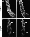
Localization of the Na,K ATPase in β spectrin mutants by confocal microscopy. Dissected preparations of the middle midgut from wild-type control larvae (A and B) and β-specem6 mutants (C and D) were stained with mouse monoclonal anti–Na,K ATPase α subunit and fluorescent secondary antibody. The basolateral staining pattern of the Na,K ATPase in wild-type appeared as rings in en face views (A), or as horseshoe shapes in optical sections near the center of the midgut (B). Neither pattern was detectable in the β-specem6 mutants (C and D). Toward the anterior (C and D, arrowhead), Na,K ATPase staining was distributed throughout the cytoplasm. Toward the posterior (C and D, arrow), staining appeared as large puncta and did not appear to be associated with the plasma membrane. In between these two regions, staining was relatively weak or in some cases absent. Bar, 10 μM.
Figure 5
Figure 5
Localization of α and βH spectrin in β spectrin mutants by confocal microscopy. Dissected preparations of middle midgut were double-labeled with mouse monoclonal anti–α spectrin (A and D) and rabbit anti–βH spectrin (B and E) and fluorescent secondary antibodies as in Fig. 3. The two subunits colocalized in the apical membrane domain of copper cells in wild-type (A–C, arrows) and in β-specem6 copper cells (D and E, arrows). Although α spectrin was most prominently visible at the basolateral surface of wild-type copper cells (A), staining of the apical surface predominated in the β spectrin mutants (D). In some cases, α and βH spectrin colocalized at the basolateral surface of mutant copper cells (D and E, arrowheads). Bar, 20 μM.
Figure 7
Figure 7
Stomach acidification was analyzed by feeding β spectrin mutant larvae and their wild-type siblings with bromphenol blue–dyed yeast paste. Acidification was scored by examining the dye color in dissected midguts. Results are presented as the percentage of larvae with strong (yellow), moderate (green), or no detectable midgut acidification (blue).
Figure 8
Figure 8
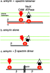
Schematic models for the flow of positional information through the spectrin membrane skeleton. (a) The spectrin tetramer includes two ankyrin binding sites that provide potential interaction sites for a source of positional information (e.g., a cell adhesion molecule [CAM]) and for other membrane markers such as the Na,K ATPase. (b) Biochemical studies indicate that mammalian ankyrin can simultaneously interact with two different integral membrane proteins. The results shown here indicate that ankyrin alone, in the absence of β spectrin, is not sufficient to direct the normal basolateral accumulation of Na,K ATPase. β spectrin may indirectly affect the activity of ankyrin, perhaps through an allosteric mechanism. (c) Alternatively, β spectrin dimers may couple two ankyrin molecules that independently associate with a source and a target of positional information. One possible mechanism of β spectrin dimer formation is through its NH2-terminal actin binding activity.

Similar articles

Cited by

References

    1. Baumann O., Lautenschlager B., Takeyasu K. Immunolocalization of Na,K-ATPase in blowfly photoreceptor cells. Cell Tissue Res. 1994;275:225–234. - PubMed
    1. Bennett V., Gilligan D.M. The spectrin-based membrane skeleton and micron-scale organization of the plasma membrane. Annu. Rev. Cell Biol. 1993;9:27–66. - PubMed
    1. Bloch R.J., Morrow J.S. An unusual beta-spectrin associated with clustered acetylcholine receptors. J. Cell Biol. 1989;108:481–493. - PMC - PubMed
    1. Byers T.J., Husain-Chishti A., Dubreuil R.R., Branton D., Goldstein L.S.B. Sequence similarity of the amino-terminal domain of Drosophila beta spectrin to alpha actinin and dystrophin. J. Cell Biol. 1989;109:1633–1641. - PMC - PubMed
    1. Byers T.J., Brandin E., Winograd E., Lue R., Branton D. The complete sequence of Drosophila beta spectrin reveals supra-motifs comprising eight 106-residue segments. Proc. Natl. Acad. Sci. USA. 1992;89:6187–6191. - PMC - PubMed

Publication types