. 2000 May 1;149(3):657-66.
doi: 10.1083/jcb.149.3.657.

Calcineurin activity is required for the initiation of skeletal muscle differentiation

Affiliations

Calcineurin activity is required for the initiation of skeletal muscle differentiation

B B Friday et al. J Cell Biol. .

Abstract

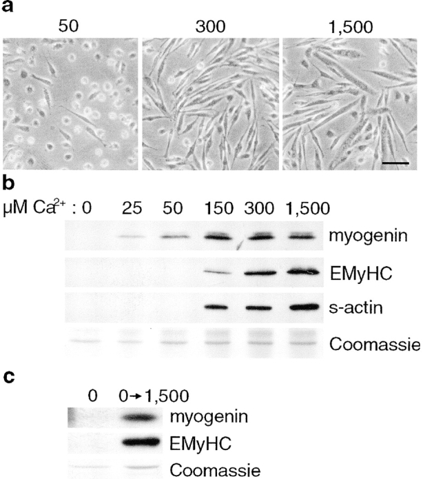
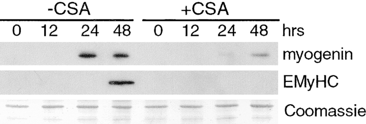
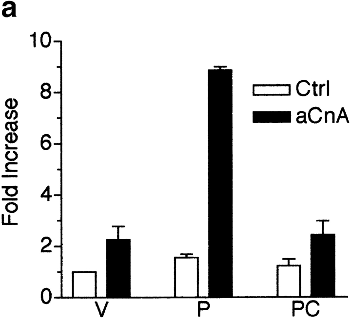
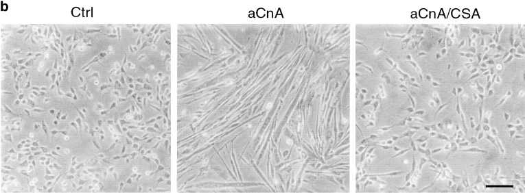
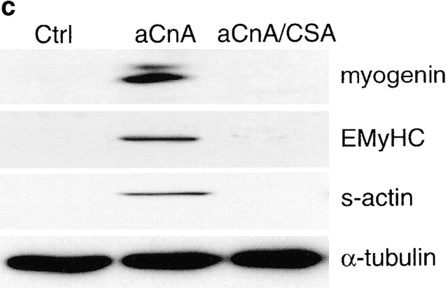
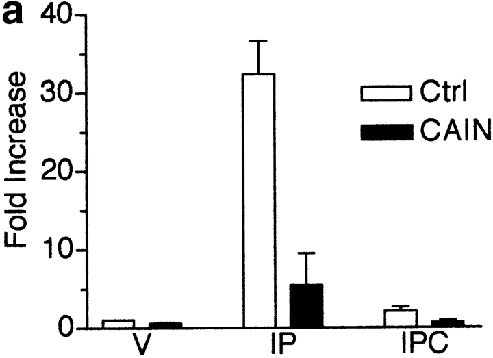
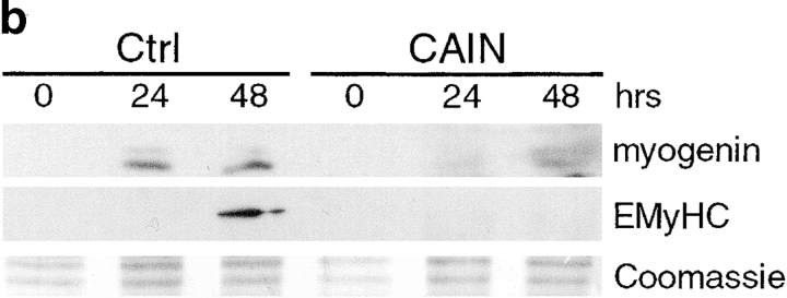
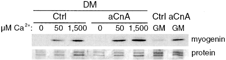
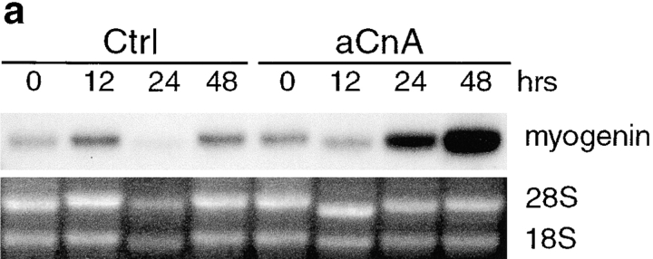
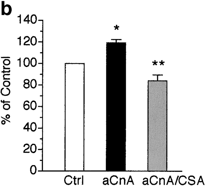
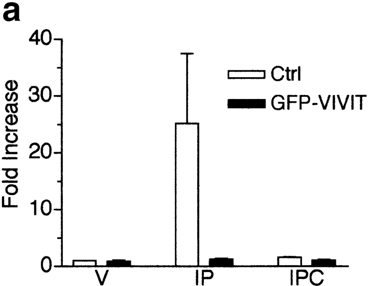
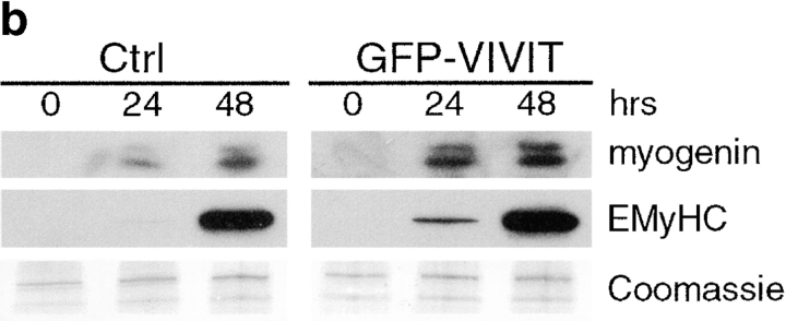
Differentiation of skeletal muscle myoblasts follows an ordered sequence of events: commitment, cell cycle withdrawal, phenotypic differentiation, and finally cell fusion to form multinucleated myotubes. The molecular signaling pathways that regulate the progression are not well understood. Here we investigate the potential role of calcium and the calcium-dependent phosphatase calcineurin in myogenesis. Commitment, phenotypic differentiation, and cell fusion are identified as distinct calcium-regulated steps, based on the extracellular calcium concentration required for the expression of morphological and biochemical markers specific to each of these stages. Furthermore, differentiation is inhibited at the commitment stage by either treatment with the calcineurin inhibitor cyclosporine A (CSA) or expression of CAIN, a physiological inhibitor of calcineurin. Retroviral-mediated gene transfer of a constitutively active form of calcineurin is able to induce myogenesis only in the presence of extracellular calcium, suggesting that multiple calcium-dependent pathways are required for differentiation. The mechanism by which calcineurin initiates differentiation includes transcriptional activation of myogenin, but does not require the participation of NFAT. We conclude that commitment of skeletal muscle cells to differentiation is calcium and calcineurin-dependent, but NFAT-independent.


Figures

Figure 1
Figure 1
Extracellular calcium is required for myoblast fusion and the expression of markers of myogenic commitment and differentiation. Primary myoblasts were induced to differentiate in DM-CF containing the indicated concentrations of exogenously added calcium for 24 h. (a) Phase-contrast images of cells grown in 50, 300, and 1,500 μM added calcium. Mononucleated cells are seen at both 50 and 300 μM Ca2+, but the cells become elongated and align at 300 μM. At 1,500 μM Ca2+, mononucleated cells fuse into multinucleated myotubes. (b) Immunoblots were performed using antibodies against markers of distinct myogenic phases. Extracellular calcium is required for significant expression of all the markers tested. A portion of a Coomassie-stained gel demonstrates relative protein loading. These results are representative of three independent experiments. (c) After 24 h in DM-CF containing no exogenously added calcium, cells were either maintained in the same media or changed to DM-CF containing 1,500 μM Ca2+ for an additional 24 h. Immunoblots demonstrate that the cells retain the ability to differentiate after the readdition of calcium. A portion of a Coomassie-stained gel demonstrates relative protein loading. Bar, 30 μm.
Figure 2
Figure 2
CSA inhibits myogenic commitment and differentiation. L6 myoblasts were induced to differentiate in DM containing either vehicle or 1 μM CSA. Cellular proteins were collected at various time points and analyzed for expression of myogenic markers. In control cells, expression of myogenin and EMyHC is first detected at 24 and 48 h, respectively. In CSA-treated cells, the expression of myogenin is decreased, and EMyHC is undetectable. A portion of a Coomassie-stained gel demonstrates relative protein loading.
Figure 4
Figure 4
A constitutively active form of calcineurin induces myogenic differentiation in primary myoblasts under growth promoting conditions. (a) Primary myoblasts containing an NFAT responsive reporter construct were infected with either control (Ctrl) or aCnA retroviruses. After 48 h, the cells were induced to differentiate by changing to DM for 24 h. Luciferase assays were performed on vehicle- (V) or PMA- (P, 10 nM) treated cultures. Some cultures were pretreated with 1 μM CSA. aCnA expression increases luciferase levels compared with control cells in PMA-treated cultures. aCnA is sensitive to inhibition by CSA. Data are reported as the fold increase over vehicle-treated control cells. Each bar represents the mean ± SEM of three independent experiments each performed in duplicate. (b and c) Primary myoblasts underwent two rounds of infection with either control (Ctrl) or aCnA retroviruses. CSA (1 μM) was added to some cultures after the second infection. (b) Phase-contrast images of cells 48 h after infection. aCnA induces morphologic changes indicative of myogenic differentiation, including cell elongation, cell alignment, and occasional cell fusion. The morphologic changes are blocked by the addition of CSA to the media. (c) 48 h after infection, proteins were collected and analyzed by immunoblotting for markers of commitment and differentiation. Expression of aCnA results in induction of multiple myogenic markers, which can be blocked with CSA. The expression of the nonmyogenic protein α-tubulin demonstrates equivalent loading of proteins. These results are representative of three independent experiments. Bar, 30 μm.
Figure 4
Figure 4
A constitutively active form of calcineurin induces myogenic differentiation in primary myoblasts under growth promoting conditions. (a) Primary myoblasts containing an NFAT responsive reporter construct were infected with either control (Ctrl) or aCnA retroviruses. After 48 h, the cells were induced to differentiate by changing to DM for 24 h. Luciferase assays were performed on vehicle- (V) or PMA- (P, 10 nM) treated cultures. Some cultures were pretreated with 1 μM CSA. aCnA expression increases luciferase levels compared with control cells in PMA-treated cultures. aCnA is sensitive to inhibition by CSA. Data are reported as the fold increase over vehicle-treated control cells. Each bar represents the mean ± SEM of three independent experiments each performed in duplicate. (b and c) Primary myoblasts underwent two rounds of infection with either control (Ctrl) or aCnA retroviruses. CSA (1 μM) was added to some cultures after the second infection. (b) Phase-contrast images of cells 48 h after infection. aCnA induces morphologic changes indicative of myogenic differentiation, including cell elongation, cell alignment, and occasional cell fusion. The morphologic changes are blocked by the addition of CSA to the media. (c) 48 h after infection, proteins were collected and analyzed by immunoblotting for markers of commitment and differentiation. Expression of aCnA results in induction of multiple myogenic markers, which can be blocked with CSA. The expression of the nonmyogenic protein α-tubulin demonstrates equivalent loading of proteins. These results are representative of three independent experiments. Bar, 30 μm.
Figure 3
Figure 3
Inhibition of calcineurin activity by CAIN inhibits myogenesis in L6 muscle cell cultures. (a) L6 myoblasts were infected with an NFAT reporter and either control (Ctrl) or CAIN retroviruses. The cells were induced to differentiate in DM for 48 h before drug treatment. Luciferase assays were performed on vehicle (V), ionomycin (I, 1 μM) plus PMA- (P, 10 nM), or IP plus CSA- (C, 1 μM) treated cultures. CAIN inhibits calcineurin in skeletal muscle cultures based on the reduction in luciferase activity in IP-treated cultures. Data are reported as the fold increase over vehicle-treated control cells. Each bar represents the mean ± SEM of three independent experiments each performed in triplicate. (b) L6 myoblasts were infected with either control or CAIN expression retroviruses and induced to differentiate in DM. Cellular proteins were collected at various time points and analyzed by immunoblotting. CAIN reduces the expression level of both myogenic markers. A portion of a Coomassie-stained membrane demonstrates relative loading of proteins. The blot shown is representative of two independent experiments.
Figure 3
Figure 3
Inhibition of calcineurin activity by CAIN inhibits myogenesis in L6 muscle cell cultures. (a) L6 myoblasts were infected with an NFAT reporter and either control (Ctrl) or CAIN retroviruses. The cells were induced to differentiate in DM for 48 h before drug treatment. Luciferase assays were performed on vehicle (V), ionomycin (I, 1 μM) plus PMA- (P, 10 nM), or IP plus CSA- (C, 1 μM) treated cultures. CAIN inhibits calcineurin in skeletal muscle cultures based on the reduction in luciferase activity in IP-treated cultures. Data are reported as the fold increase over vehicle-treated control cells. Each bar represents the mean ± SEM of three independent experiments each performed in triplicate. (b) L6 myoblasts were infected with either control or CAIN expression retroviruses and induced to differentiate in DM. Cellular proteins were collected at various time points and analyzed by immunoblotting. CAIN reduces the expression level of both myogenic markers. A portion of a Coomassie-stained membrane demonstrates relative loading of proteins. The blot shown is representative of two independent experiments.
Figure 5
Figure 5
Multiple calcium-dependent pathways are necessary for initiating myogenesis. Myoblasts were infected with either control (Ctrl) or aCnA retroviruses. Cells were maintained in GM for 6 h and then induced to differentiate in DM-CF containing different concentrations of exogenously added calcium for 36 h. Some cultures were kept in GM for the entire experiment. Cellular proteins were analyzed by immunoblotting using an antibody against myogenin. aCnA-infected cells demonstrate a calcium-dependent expression pattern of myogenin similar to control cells. The absence of the expression of myogenin at 0 μM Ca 2+ is not due to inefficient infection of the cells, because aCnA-infected cells maintained in GM express myogenin at higher levels than control cells. A portion of a membrane stained for total protein with colloidal gold is shown to indicate relative protein loading. The data are representative of three independent experiments.
Figure 6
Figure 6
aCnA increases levels of myogenin mRNA and activates transcription from a myogenin reporter construct. (a) Primary myoblasts were infected with either control (Ctrl) or aCnA retroviruses and maintained in GM. RNA samples were collected at various time points and analyzed by Northern blotting for myogenin mRNA. Expression of aCnA results in an increase in myogenin mRNA compared with control cultures. A portion of an ethidium bromide–stained gel containing 28S and 18S rRNAs demonstrates relative RNA loading. The blot shown is representative of two independent experiments. (b) Primary myoblasts containing a myogenin promoter reporter construct were infected with either control or aCnA retroviruses. After 48 h in GM, the cultures were analyzed for luciferase activity. Expression of aCnA results in a CSA-sensitive activation of the myogenin promoter. Each bar represents the mean ± SEM of 3–4 independent experiments each performed in triplicate. Statistical analyses were performed using one-way ANOVA with Bonferroni's multiple comparison test. *P < 0.01 vs Ctrl; **P < 0.001 vs aCnA.
Figure 6
Figure 6
aCnA increases levels of myogenin mRNA and activates transcription from a myogenin reporter construct. (a) Primary myoblasts were infected with either control (Ctrl) or aCnA retroviruses and maintained in GM. RNA samples were collected at various time points and analyzed by Northern blotting for myogenin mRNA. Expression of aCnA results in an increase in myogenin mRNA compared with control cultures. A portion of an ethidium bromide–stained gel containing 28S and 18S rRNAs demonstrates relative RNA loading. The blot shown is representative of two independent experiments. (b) Primary myoblasts containing a myogenin promoter reporter construct were infected with either control or aCnA retroviruses. After 48 h in GM, the cultures were analyzed for luciferase activity. Expression of aCnA results in a CSA-sensitive activation of the myogenin promoter. Each bar represents the mean ± SEM of 3–4 independent experiments each performed in triplicate. Statistical analyses were performed using one-way ANOVA with Bonferroni's multiple comparison test. *P < 0.01 vs Ctrl; **P < 0.001 vs aCnA.
Figure 7
Figure 7
Inhibition of NFAT activity by the expression of GFP-VIVIT does not inhibit myogenesis in L6 muscle cell cultures. (a) Primary myoblasts were infected with an NFAT responsive reporter and either control (Ctrl) or GFP-VIVIT retroviruses. Cells were induced to differentiate for 48 h in DM. Luciferase assays were performed on vehicle (V), ionomycin (I, 1 μM) plus PMA- (P, 10 nM), or IP plus CSA- (C, 1 μM) treated cultures. GFP-VIVIT inhibits NFAT activity in skeletal muscle cells. Data are reported as the fold increase over vehicle-treated control cells. Each bar represents the mean ± SEM of three independent experiments each performed in triplicate. (b) L6 myoblasts were infected with either control or GFP-VIVIT retroviruses and induced to differentiate in DM. Cellular proteins were collected at various time points and analyzed by immunoblotting. GFP-VIVIT does not inhibit the expression of either myogenic marker. The blot shown is representative of two independent experiments.
Figure 7
Figure 7
Inhibition of NFAT activity by the expression of GFP-VIVIT does not inhibit myogenesis in L6 muscle cell cultures. (a) Primary myoblasts were infected with an NFAT responsive reporter and either control (Ctrl) or GFP-VIVIT retroviruses. Cells were induced to differentiate for 48 h in DM. Luciferase assays were performed on vehicle (V), ionomycin (I, 1 μM) plus PMA- (P, 10 nM), or IP plus CSA- (C, 1 μM) treated cultures. GFP-VIVIT inhibits NFAT activity in skeletal muscle cells. Data are reported as the fold increase over vehicle-treated control cells. Each bar represents the mean ± SEM of three independent experiments each performed in triplicate. (b) L6 myoblasts were infected with either control or GFP-VIVIT retroviruses and induced to differentiate in DM. Cellular proteins were collected at various time points and analyzed by immunoblotting. GFP-VIVIT does not inhibit the expression of either myogenic marker. The blot shown is representative of two independent experiments.

References

    1. Abbott K.L., Friday B.B., Thaloor D., Murphy T.J., Pavlath G.K. Activation and cellular localization of the cyclosporine A-sensitive transcription factor NF-AT in skeletal muscle cells. Mol. Biol. Cell. 1998;9:2905–2916. - PMC - PubMed
    1. Andres V., Walsh K. Myogenin expression, cell cycle withdrawal, and phenotypic differentiation are temporally separable events that precede cell fusion upon myogenesis. J. Cell Biol. 1996;132:657–666. - PMC - PubMed
    1. Aramburu J., Yaffe M.B., Lopez-Rodriguez C., Cantley L.C., Hogan P.G., Rao A. Affinity-driven peptide selection of an NFAT inhibitor more selective than cyclosporin A. Science. 1999;285:2129–2133. - PubMed
    1. Bar-Sagi D., Prives J. Trifluoperazine, a calmodulin antagonist, inhibits muscle cell fusion. J. Cell Biol. 1983;97:1375–1380. - PMC - PubMed
    1. Barnoy S., Glaser T., Kosower N.S. Calpain and calpastatin in myoblast differentiation and fusioneffects of inhibitors. Biochim. Biophys. Acta. 1997;1358:181–188. - PubMed

Publication types

MeSH terms