. 2000 May 1;149(3):697-706.
doi: 10.1083/jcb.149.3.697.

Focal exocytosis of VAMP3-containing vesicles at sites of phagosome formation

Affiliations

Focal exocytosis of VAMP3-containing vesicles at sites of phagosome formation

L Bajno et al. J Cell Biol. .

Abstract

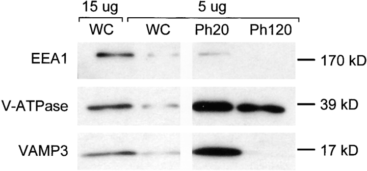
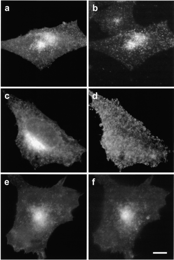
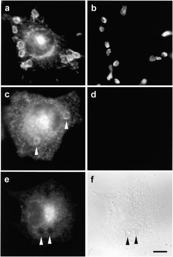
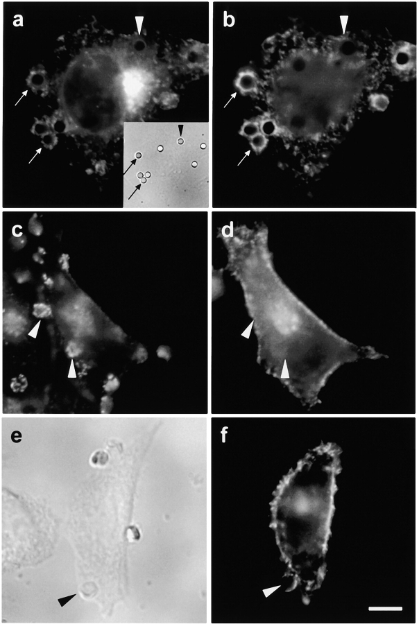
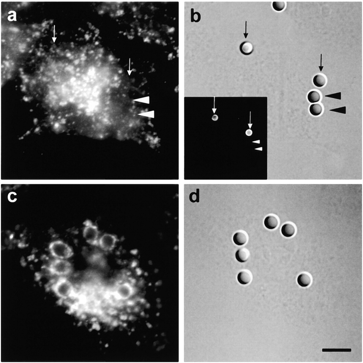
Phagocytosis involves the receptor-mediated extension of plasmalemmal protrusions, called pseudopods, which fuse at their tip to engulf a particle. Actin polymerizes under the nascent phagosome and may propel the protrusion of pseudopods. Alternatively, membrane extension could result from the localized insertion of intracellular membranes into the plasmalemma next to the particle. Here we show focal accumulation of VAMP3-containing vesicles, likely derived from recycling endosomes, in the vicinity of the nascent phagosome. Using green fluorescent protein (GFP) as both a fluorescent indicator and an exofacial epitope tag, we show that polarized fusion of VAMP3 vesicles precedes phagosome sealing. It is therefore likely that targeted delivery of endomembranes contributes to the elongation of pseudopods. In addition to mediating pseudopod formation, receptor-triggered focal secretion of endosomes may contribute to polarized membrane extension in processes such as lamellipodial elongation or chemotaxis.


Figures

Figure 1
Figure 1
VAMP3 is enriched in early phagosomes. J774 cells were allowed to internalize latex beads for either 20 min or 2 h and phagosomes were purified by gradient centrifugation. Samples of whole cell lysates (WC) or of phagosomes formed after 20 min (Ph20) or 2 h (Ph120) were resolved by SDS-PAGE and immunoblotted with polyclonal antibodies against EEA1 (top), the 39-kD subunit of the V-ATPase (middle), and VAMP3 (bottom). The amount of protein loaded in each lane is indicated at the top. Representative of at least three similar experiments.
Figure 2
Figure 2
Localization and recycling of VAMP3 in CHO-IIA cells. (a and b) CHO-IIA cells transfected transiently with VAMP3-GFP were incubated with 25 μg/ml tetramethylrhodamine-labeled transferrin for 1 h at 37°C, washed and visualized by fluorescence microscopy. (a) VAMP3-GFP (green fluorescence); (b) transferrin (red fluorescence). (c–f) CHO-IIA cells transiently cotransfected with VAMP3-GFP and VAMP3-Ig were incubated at 4°C with Cy3-labeled F(ab) fragments of antibodies raised against human IgG, to label exofacial VAMP3-Ig. In c and d the cells were visualized at this stage. In e and f, the cells were next warmed to 37°C and incubated for an additional h to allow internalization of the labeled VAMP3-Ig. (c and e) VAMP3-GFP (green fluorescence); (d and f) Cy3-labeled VAMP-Ig (red fluorescence). Representative of at least three similar experiments. Bar, 10 μm.
Figure 3
Figure 3
Localization of VAMP3-GFP in CHO-IIA cells during phagocytosis. CHO-IIA cells transfected with VAMP3-GFP were incubated at 37°C with RBC-Ig for 10 (a and b), 30 (c and d), or 90 min (e and f). The distribution of VAMP3-GFP (green fluorescence) is shown in a, c, and e. In b and d, extracellular RBC-Ig were detected by incubation at 4°C with Cy3-labeled antibodies against the opsonizing IgG. f shows the differential interference contrast image of the cell in e. Arrowheads indicate internalized RBC-Ig. Representative of at least three similar experiments of each type. Bar, 10 μm.
Figure 4
Figure 4
Surface exposure of VAMP3-GFP in CHO-IIA cells during phagocytosis. (a–b) CHO-IIA cells transfected with VAMP3-GFP were incubated at 37°C with IgG-opsonized latex beads for 10 min. After stopping phagocytosis by cooling to 4°C, exofacial VAMP3-GFP was detected by incubation with antibodies to GFP followed by secondary Cy3-conjugated antibodies. (a) Distribution of VAMP3-GFP (green fluorescence). Arrows indicate beads that are undergoing phagocytosis, whereas the arrowhead indicates a bead not being internalized. Inset shows the corresponding bright field image. (b) Distribution of exofacial VAMP3-GFP (red fluorescence). (c and d) Cells stably expressing FcγRIIA-GFP were transiently transfected with VAMP3-Ig and then incubated at 4°C with Cy3-labeled F(ab) fragments of antibodies raised against human IgG, to label exofacial VAMP3-Ig. Finally, opsonized RBC were added to induce receptor clustering. (d) Distribution of FcγRIIA-GFP (green fluorescence). Arrowheads point to FcγRIIA-GFP clusters formed at sites of interaction with the RBC. (d) Distribution of VAMP3-Ig (red fluorescence). (e and f) CHO-IIA cells were transiently transfected with PM-GFP. Next, RBC-Ig were added to initiate phagosome formation. (e) Bright field image. Arrowhead indicates a nascent phagosome. (f) Distribution of PM-GFP cell in (e). Representative of at least three experiments of each type. Bar, 10 μm.
Figure 5
Figure 5
Localization of LAMP1 in CHO-IIA cells during phagocytosis. CHO-IIA cells were incubated at 37°C with IgG-opsonized latex beads for 10 min (a and b) or 60 min (c and d). The distribution of LAMP1, detected by indirect immunofluorescence using Cy3-labeled antibodies, is shown in a and c. b and d show the differential interference contrast images of the cells in a and c, respectively. In a and b, arrowheads indicate beads that have been internalized, whereas arrows indicate extracellular beads, identified by accessibility to extracellularly added labeled antibodies. (Inset) Extracellular particles were detected by incubation at 4°C with FITC-labeled antibodies against the opsonizing IgG. Bar, 10 μm (does not apply to inset).

Comment in

Similar articles

Cited by

References

    1. Adams A.E., Johnson D.I., Longnecker R.M., Sloat B.F., Pringle J.R. CDC42 and CDC43, two additional genes involved in budding and the establishment of cell polarity in the yeast Saccharomyces cerevisiae . J. Cell Biol. 1990;111:131–142. - PMC - PubMed
    1. Betz W.J., Bewick G.S. Optical analysis of synaptic vesicle recycling at the frog neuromuscular junction. Science. 1992;255:200–203. - PubMed
    1. Bretscher M.S. Getting membrane flow and the cytoskeleton to cooperate in moving cells. Cell. 1996;87:601–606. - PubMed
    1. Buys S.S., Keogh E.A., Kaplan J. Fusion of intracellular membrane pools with cell surfaces of macrophages stimulated by phorbol esters and calcium ionophores. Cell. 1984;38:569–576. - PubMed
    1. Cox D., Tseng C.C., Bjekic G., Greenberg S. A requirement for phosphatidylinositol 3-kinase in pseudopod extension. J. Biol. Chem. 1999;274:1240–1247. - PubMed

Publication types

MeSH terms