. 2000 May 1;149(3):741-54.
doi: 10.1083/jcb.149.3.741.

Matrix survival signaling: from fibronectin via focal adhesion kinase to c-Jun NH(2)-terminal kinase

Affiliations

Matrix survival signaling: from fibronectin via focal adhesion kinase to c-Jun NH(2)-terminal kinase

E A Almeida et al. J Cell Biol. .

Abstract

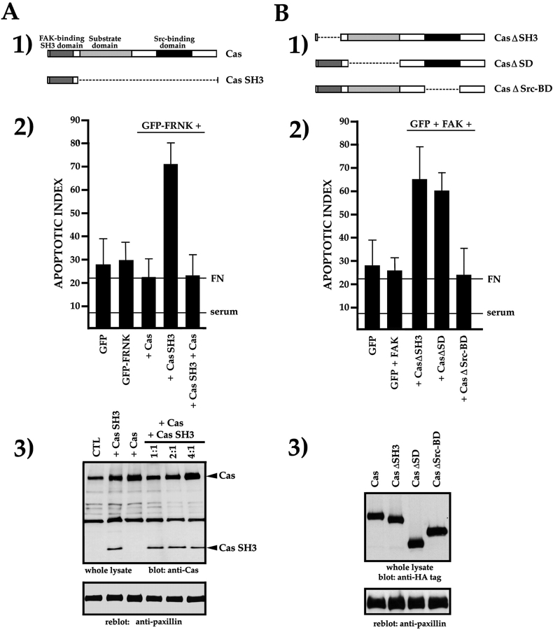
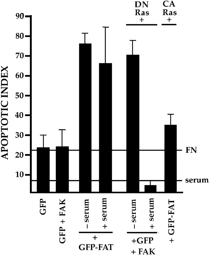
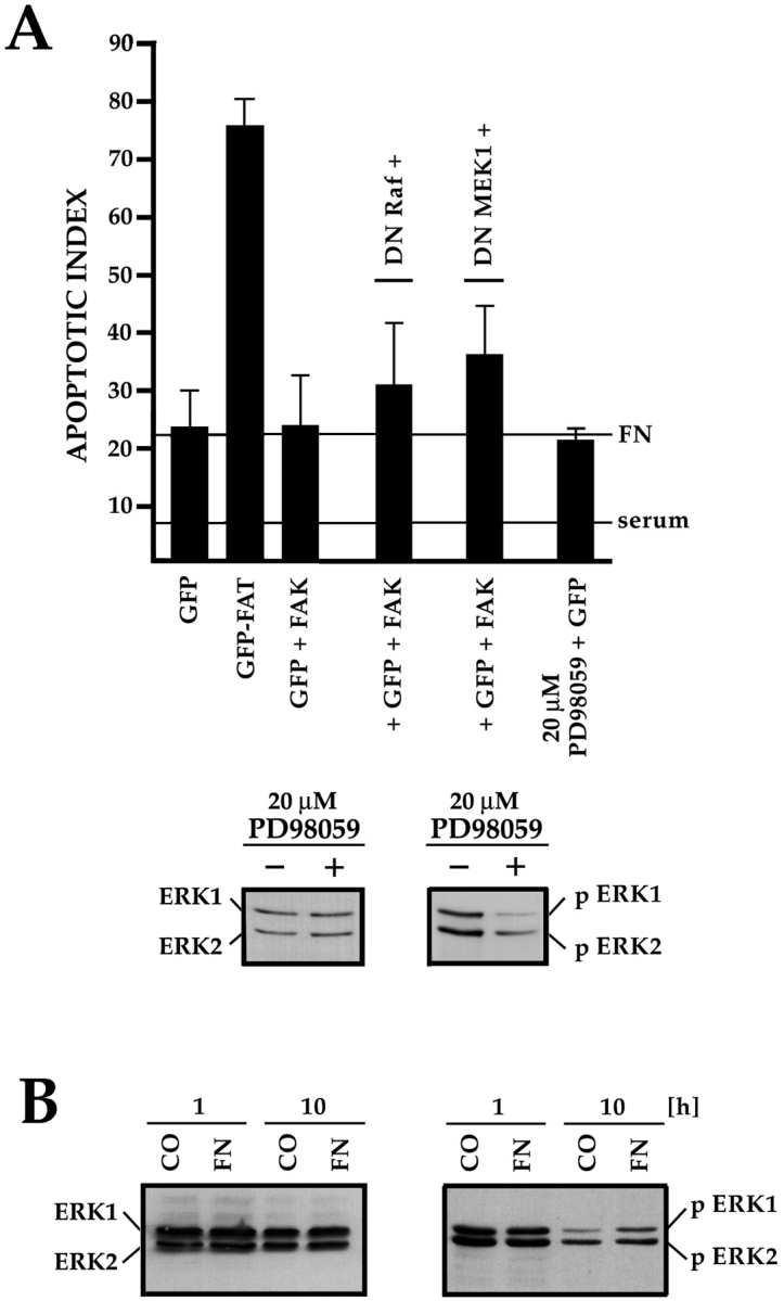
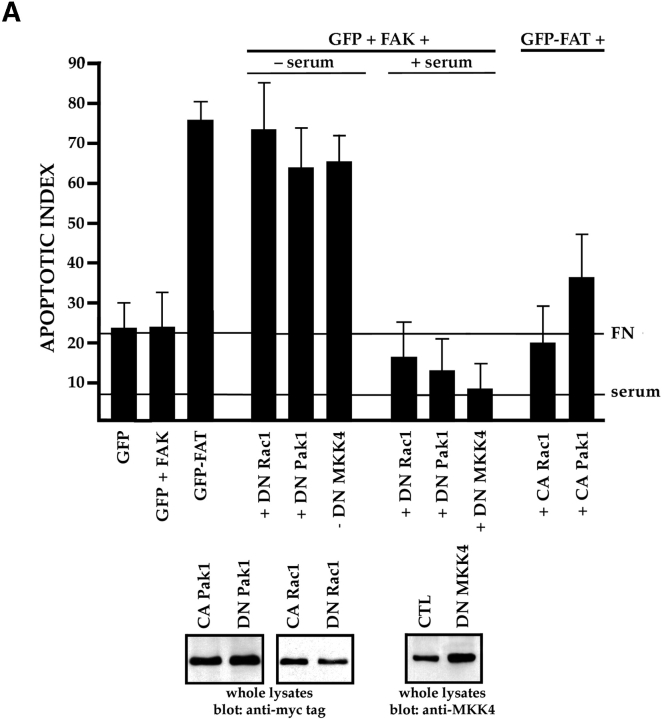
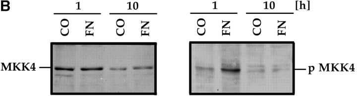
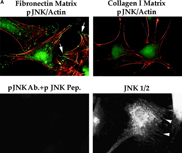
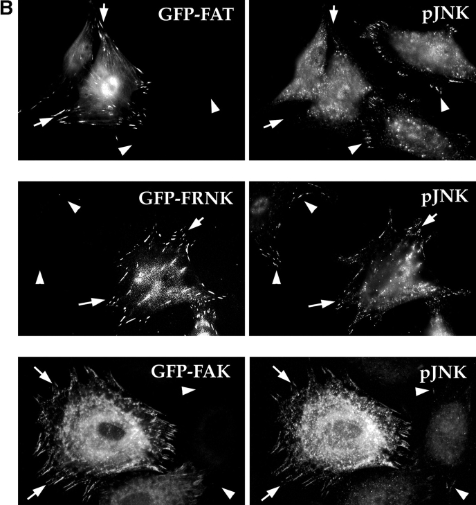
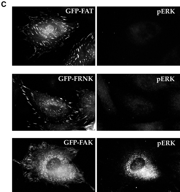
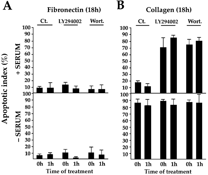
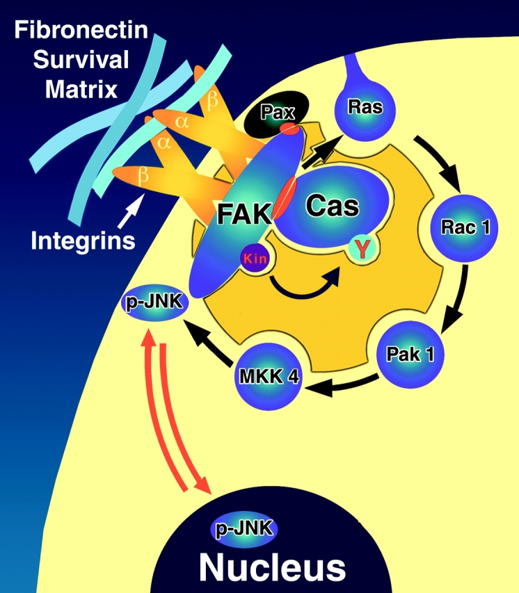
Most transformed cells have lost anchorage and serum dependence for growth and survival. Previously, we established that when serum is absent, fibronectin survival signals transduced by focal adhesion kinase (FAK), suppress p53-regulated apoptosis in primary fibroblasts and endothelial cells (Ilić et al. 1998. J. Cell Biol. 143:547-560). The present goals are to identify survival sequences in FAK and signaling molecules downstream of FAK required for anchorage-dependent survival of primary fibroblasts. We report that binding of the SH3 domain of p130Cas to proline-rich region 1 of FAK is required to support survival of fibroblasts on fibronectin when serum is withdrawn. The FAK-p130Cas complex activates c-Jun NH2-terminal kinase (JNK) via a Ras/Rac1/Pak1/MAPK kinase 4 (MKK4) pathway. Activated (phospho-) JNK colocalizes with FAK in focal adhesions of fibroblasts cultured on fibronectin, which supports their survival, but not in fibroblasts cultured on collagen, which does not. Cells often survive in the absence of extracellular matrix if serum factors are provided. In that case, we confirm work of others that survival signals are transduced by FAK, phosphatidylinositol 3'-kinase (PI3-kinase), and Akt/protein kinase B (PKB). However, when serum is absent, PI3-kinase and Akt/PKB are not involved in the fibronectin-FAK-JNK survival pathway documented herein. Thus, survival signals from extracellular matrix and serum are transduced by FAK via two distinct pathways.


Figures

Figure 1
Figure 1
The PR-1 region of FAK/FRNK and focal contact localization are required for the transmission of FN matrix survival signals. RSF plated on FN in the presence of serum were transfected with wild-type FAK and GFP, GFP-FRNK, GFP-FAT, or GFP-FRNK with mutations in PR-1. Cells were cultured subsequently in the absence of serum for the times indicated. The apoptotic index was assessed by determining the percentage of transfected cells (GFP-positive) showing condensed or fragmented nuclei after staining with Hoechst dye. (A) Cells expressing GFP+FAK for 16 h remain spread on a FN matrix (upper left panel). Cells expressing GFP-FAT for 16 h (upper right panel) are rounded with condensed, fragmented nuclei. In cells expressing GFP-FAT for 8, 12, and 16 h (lower left panel), GFP-FAT initially localizes to focal contacts in spread cells. By 12 h, cells expressing GFP-FAT start to round. By 16 h, rounded cells have condensed nuclei as detected by Hoechst staining (arrow). Adjacent cell not expressing GFP-FAT has a normal nucleus (indicated by asterisk). In cells expressing GFP-FRNK for 16 h (lower right panel), GFP-FRNK localizes to focal contacts, but cells survive. (B) Expression levels of FAK, GFP-FRNK, and GFP-FAT in RSF 8 h after transfection. All are expressed at similar levels as shown by Western blot using a COOH-terminal FAK antibody. (C) GFP-FAT (upper row) and GFP-FRNK (lower row) both replace endogenous FAK in focal contacts. GFP, but not endogenous FAK, which is detected by an antibody against the NH2-terminal domain of FAK (N-FAK), is present in focal contacts of transfected cells (arrowheads). Nontransfected (GFP-negative) cells in the same field stain with N-FAK (arrows). (D) Diagrammatic representation of constructs used to test the importance of PR-1 in FAK and FRNK for supporting survival of RSF on a FN matrix (see text for description). (E) Apoptotic index for RSF transfected with constructs shown in D after 16 h. Mutation of PR-1 in both FRNK and FAK promotes apoptosis at levels comparable to GFP-FAT. A GFP-FRNK mutant that does not localize to focal contacts but has intact prolines 712–713 (GFP-FRNKΔFAT) is pro-apoptotic, whereas the GFP-FRNK P mutant that does not localize to focal contacts (GFP-FRNK PΔFAT) loses the ability to induce apoptosis. PBD, paxillin-binding domain.
Figure 1
Figure 1
The PR-1 region of FAK/FRNK and focal contact localization are required for the transmission of FN matrix survival signals. RSF plated on FN in the presence of serum were transfected with wild-type FAK and GFP, GFP-FRNK, GFP-FAT, or GFP-FRNK with mutations in PR-1. Cells were cultured subsequently in the absence of serum for the times indicated. The apoptotic index was assessed by determining the percentage of transfected cells (GFP-positive) showing condensed or fragmented nuclei after staining with Hoechst dye. (A) Cells expressing GFP+FAK for 16 h remain spread on a FN matrix (upper left panel). Cells expressing GFP-FAT for 16 h (upper right panel) are rounded with condensed, fragmented nuclei. In cells expressing GFP-FAT for 8, 12, and 16 h (lower left panel), GFP-FAT initially localizes to focal contacts in spread cells. By 12 h, cells expressing GFP-FAT start to round. By 16 h, rounded cells have condensed nuclei as detected by Hoechst staining (arrow). Adjacent cell not expressing GFP-FAT has a normal nucleus (indicated by asterisk). In cells expressing GFP-FRNK for 16 h (lower right panel), GFP-FRNK localizes to focal contacts, but cells survive. (B) Expression levels of FAK, GFP-FRNK, and GFP-FAT in RSF 8 h after transfection. All are expressed at similar levels as shown by Western blot using a COOH-terminal FAK antibody. (C) GFP-FAT (upper row) and GFP-FRNK (lower row) both replace endogenous FAK in focal contacts. GFP, but not endogenous FAK, which is detected by an antibody against the NH2-terminal domain of FAK (N-FAK), is present in focal contacts of transfected cells (arrowheads). Nontransfected (GFP-negative) cells in the same field stain with N-FAK (arrows). (D) Diagrammatic representation of constructs used to test the importance of PR-1 in FAK and FRNK for supporting survival of RSF on a FN matrix (see text for description). (E) Apoptotic index for RSF transfected with constructs shown in D after 16 h. Mutation of PR-1 in both FRNK and FAK promotes apoptosis at levels comparable to GFP-FAT. A GFP-FRNK mutant that does not localize to focal contacts but has intact prolines 712–713 (GFP-FRNKΔFAT) is pro-apoptotic, whereas the GFP-FRNK P mutant that does not localize to focal contacts (GFP-FRNK PΔFAT) loses the ability to induce apoptosis. PBD, paxillin-binding domain.
Figure 2
Figure 2
The Cas SH3 and SD are required for transmission of survival signals from FN and FAK. (A, panel 1) Diagram of the domain structure of Cas and the isolated Cas SH3 domain. (A, panel 2) Expression of isolated Cas SH3 domain with GFP-FRNK disrupts survival. Coexpression of full-length Cas and Cas SH3 in a 4:1 DNA molar ratio restores survival. (A, panel 3) Western blot of lysates of cells transfected with Cas SH3 or Cas, and with both Cas and Cas SH3 at increasing ratios of Cas cDNA. A paxillin reblot of the same membrane shows that sample loading is similar in all lanes. (B, panel 1) Diagrams of Cas cDNAs with deletions of the SH3 domain (CasΔSH3), the substrate-binding domain (CasΔSD), or the Src-binding domain (CasΔSrc-BD). (B, panel 2) Expression of CasΔSH3 or CasΔSD triggers high levels of apoptosis, whereas expression of CasΔSrcBD does not. (B, panel 3) Western blot showing that all Cas constructs are expressed in RSF at similar levels. Reblot of the same membrane with an anti-paxillin antibody shows similar loading in all lanes.
Figure 3
Figure 3
Ras is involved in the transduction of matrix-dependent survival signals from FN and FAK. Expression of GFP-FAT triggered high apoptosis in RSF cultured either in the presence or absence of serum. DN Ras promoted a high level of apoptosis when coexpressed with GFP+FAK in RSF in the absence, but not in the presence of serum. Expression of constitutively active (CA) Ras rescued GFP-FAT–mediated cell death of RSF plated on FN in the absence of serum.
Figure 4
Figure 4
The ERK-MAPK pathway is not essential for supporting FN-FAK–dependent survival. (A) Cotransfection of DN Raf1 or DN MEK1, along with GFP+FAK, resulted in only a small increase in apoptosis compared with GFP+FAK alone. Addition of the MEK1/2 inhibitor PD98059 at 10× the IC50 to cultures of nontransfected RSF did not trigger elevated apoptosis. The PD98059 was active, as treated cultures (+) showed reduced levels of activated pERK1/2 compared with untreated cultures (−). Levels of total ERK proteins were similar in both cultures. (B) ERK1/2 were activated similarly by contact with FN, which supports survival, or collagen I (CO), which does not. Lysates of nontransfected RSF cultured in the absence of serum for either 1 or 10 h, were immunoblotted using antibodies that detect total ERK1/2, or phosphorylated pERK1/2.
Figure 5
Figure 5
The JNK MAPK pathway transduces survival signals from FN-FAK via Rac1/Pak1/MKK4 after serum withdrawal. (A) Expression of DN Rac1, DN Pak1, or DN MKK4, along with GFP+FAK, triggered high levels of apoptosis in RSF cultured on FN in the absence, but not in the presence of serum. Coexpression of constitutively active forms of Rac1 (CA Rac1) or Pak1 (CA Pak1) rescued cells from GFP-FAT–mediated death. The Western blot panels show that CA and DN Rac1/Pak1 construct pairs were expressed at similar levels in RSF and that DN MKK4 was expressed at a level similar to the other constructs. (B) Active MKK4 was detected in lysates of cells plated on FN but not on collagen I. Lysates of RSF cultured in the absence of serum for either 1 or 10 h on FN or on collagen I were immunoblotted using antibodies that detect total MKK4 protein or pMKK4. MKK4 protein levels were similar to one another on both matrices at each timepoint. Significant levels of pMMK4 were detected only in lysates of RSF cultured on FN at the 1 h timepoint. (C) MKK4-null and wild-type (Wt) ES cells were grown in suspension for 12 d in the presence of serum. Serum was withdrawn for 24 h and embryoid bodies were processed to detect DNA fragmentation by TUNEL in situ staining. Only MKK4-null embryoid bodies showed increased TUNEL staining when serum was withdrawn. (D) JNK, but not p38 MAPK, was activated in response to attachment to FN. Lysates of RSF cultured without serum for 1 or 10 h on FN or collagen I (CO), were immunoblotted using antibodies that detect p38 MAPK protein or pp38, or JNK1/2 proteins or pJNK1/2. p38 MAPK was only weakly activated on FN. Lysates of anisomycin-treated primary RSF were used as a positive control (CTL). The signal for pJNK2 was particularly strong at 10 h in cells plated on FN. JNK1 and JNK2 protein levels in cells on FN and collagen were similar to one another at each timepoint. (E) Active pJNK (rhodamine) is strongly visible in cells transfected with GFP-FAK (green), but remains at low levels in adjacent untransfected cells (see nuclei with no corresponding GFP staining in the merged picture). Cells on FN in the absence of serum were fixed and stained after 16 h. (F) FAK, but not GFP, activates JNK1/2 in RSF. Flag-tagged JNK1 or JNK2, and FAK or GFP were coexpressed in RSF for 16 h after serum withdrawal. Cell lysates were incubated with anti-Flag antibody to precipitate JNK1/2. Precipitates were tested for their kinase activities using glutathione S-transferase–c-Jun as a substrate. JNK1/2 were activated strongly by FAK, but not by GFP.
Figure 5
Figure 5
The JNK MAPK pathway transduces survival signals from FN-FAK via Rac1/Pak1/MKK4 after serum withdrawal. (A) Expression of DN Rac1, DN Pak1, or DN MKK4, along with GFP+FAK, triggered high levels of apoptosis in RSF cultured on FN in the absence, but not in the presence of serum. Coexpression of constitutively active forms of Rac1 (CA Rac1) or Pak1 (CA Pak1) rescued cells from GFP-FAT–mediated death. The Western blot panels show that CA and DN Rac1/Pak1 construct pairs were expressed at similar levels in RSF and that DN MKK4 was expressed at a level similar to the other constructs. (B) Active MKK4 was detected in lysates of cells plated on FN but not on collagen I. Lysates of RSF cultured in the absence of serum for either 1 or 10 h on FN or on collagen I were immunoblotted using antibodies that detect total MKK4 protein or pMKK4. MKK4 protein levels were similar to one another on both matrices at each timepoint. Significant levels of pMMK4 were detected only in lysates of RSF cultured on FN at the 1 h timepoint. (C) MKK4-null and wild-type (Wt) ES cells were grown in suspension for 12 d in the presence of serum. Serum was withdrawn for 24 h and embryoid bodies were processed to detect DNA fragmentation by TUNEL in situ staining. Only MKK4-null embryoid bodies showed increased TUNEL staining when serum was withdrawn. (D) JNK, but not p38 MAPK, was activated in response to attachment to FN. Lysates of RSF cultured without serum for 1 or 10 h on FN or collagen I (CO), were immunoblotted using antibodies that detect p38 MAPK protein or pp38, or JNK1/2 proteins or pJNK1/2. p38 MAPK was only weakly activated on FN. Lysates of anisomycin-treated primary RSF were used as a positive control (CTL). The signal for pJNK2 was particularly strong at 10 h in cells plated on FN. JNK1 and JNK2 protein levels in cells on FN and collagen were similar to one another at each timepoint. (E) Active pJNK (rhodamine) is strongly visible in cells transfected with GFP-FAK (green), but remains at low levels in adjacent untransfected cells (see nuclei with no corresponding GFP staining in the merged picture). Cells on FN in the absence of serum were fixed and stained after 16 h. (F) FAK, but not GFP, activates JNK1/2 in RSF. Flag-tagged JNK1 or JNK2, and FAK or GFP were coexpressed in RSF for 16 h after serum withdrawal. Cell lysates were incubated with anti-Flag antibody to precipitate JNK1/2. Precipitates were tested for their kinase activities using glutathione S-transferase–c-Jun as a substrate. JNK1/2 were activated strongly by FAK, but not by GFP.
Figure 6
Figure 6
(A) pJNK is detected both in focal contacts and in the nucleus in cells plated on FN, but not in cells plated on collagen I. RSF plated on FN or collagen I for 10 h were fixed, permeabilized, and stained with antibodies specific for pJNK. Actin was detected by rhodamine-phalloidin. In cells on FN, strong staining for pJNK was detected in the nucleus and in focal contact sites at the ends of actin stress fibers (arrowheads). In cells plated on collagen I, stress fibers and focal contacts were poorly developed. Staining for pJNK was weak and diffuse in the cytoplasm and nucleus. As a control, the anti-pJNK antibody was added to RSF in the presence of 100 μg/ml of the pJNK peptide used to generate the antibody. No focal contact staining was detected (lower left panel). Antibodies that recognize all forms of JNK 1/2 showed a diffuse cytoplasmic staining pattern as well as faint focal contact staining (arrowheads, lower right panel). (B) Expression of GFP-FAT (upper panels) in RSF plated on FN in serum-free medium for 8–10 h (a time before appearance of apoptotic changes; see also Fig. 1), prevents pJNK location in focal contacts (arrows). Staining for pJNK was strong in focal contact sites (arrowheads) of adjacent cells that were not transfected. Expression of GFP-FRNK (middle panels) does not bar pJNK from focal contacts (arrows). pJNK staining in focal contacts of GFP-FAT–expressing cells is similar in intensity to that in focal contacts of adjacent nontransfected cells (arrowheads). Expression of GFP-FAK (lower panels) markedly increases pJNK staining in focal contacts (arrows) as well as in the cytoplasm, relative to adjacent untransfected cells (arrowheads). (C) Staining for pERK in both GFP-FAT (upper panels) and GFP-FRNK–expressing RSF (middle panels) is weak and not detected in focal contacts. pERK staining is strong in RSF expressing GFP-FAK (lower panels). However, focal contact staining is not detected.
Figure 6
Figure 6
(A) pJNK is detected both in focal contacts and in the nucleus in cells plated on FN, but not in cells plated on collagen I. RSF plated on FN or collagen I for 10 h were fixed, permeabilized, and stained with antibodies specific for pJNK. Actin was detected by rhodamine-phalloidin. In cells on FN, strong staining for pJNK was detected in the nucleus and in focal contact sites at the ends of actin stress fibers (arrowheads). In cells plated on collagen I, stress fibers and focal contacts were poorly developed. Staining for pJNK was weak and diffuse in the cytoplasm and nucleus. As a control, the anti-pJNK antibody was added to RSF in the presence of 100 μg/ml of the pJNK peptide used to generate the antibody. No focal contact staining was detected (lower left panel). Antibodies that recognize all forms of JNK 1/2 showed a diffuse cytoplasmic staining pattern as well as faint focal contact staining (arrowheads, lower right panel). (B) Expression of GFP-FAT (upper panels) in RSF plated on FN in serum-free medium for 8–10 h (a time before appearance of apoptotic changes; see also Fig. 1), prevents pJNK location in focal contacts (arrows). Staining for pJNK was strong in focal contact sites (arrowheads) of adjacent cells that were not transfected. Expression of GFP-FRNK (middle panels) does not bar pJNK from focal contacts (arrows). pJNK staining in focal contacts of GFP-FAT–expressing cells is similar in intensity to that in focal contacts of adjacent nontransfected cells (arrowheads). Expression of GFP-FAK (lower panels) markedly increases pJNK staining in focal contacts (arrows) as well as in the cytoplasm, relative to adjacent untransfected cells (arrowheads). (C) Staining for pERK in both GFP-FAT (upper panels) and GFP-FRNK–expressing RSF (middle panels) is weak and not detected in focal contacts. pERK staining is strong in RSF expressing GFP-FAK (lower panels). However, focal contact staining is not detected.
Figure 7
Figure 7
PI3-kinase is not required for transduction of survival signals if cells are spread on a pro-survival matrix (i.e., FN) whether or not serum is present, but is required to transduce serum-dependent survival signals when a pro-survival matrix is withdrawn. (A) PI3-kinase inhibitors Ly297082 and wortmannin (Wort.) have no effect on the high survival of RSF plated on FN whether or not serum is present. Inhibitors were added to cells at the time of plating or to cultures 1 h after plating. Apoptotic index was measured 18 h later. (B) PI3-kinase inhibitors promote apoptosis of cells plated on collagen I when serum is present. These inhibitors do not alter the normally high apoptotic index of cultures plated on collagen I in the absence of serum.
Figure 8
Figure 8
Model of the FN-dependent matrix survival pathway activated in primary RSF when serum is absent. FN supports the organization of a pro-survival signaling complex based on a FAK/Cas scaffold located at focal adhesion sites. Paxillin (Pax) is thought to participate in the binding of FAK to focal adhesion sites (Tachibana et al. 1995). Cas is recruited to the survival complex via a FAK PR-1–Cas SH3 interaction. Matrix survival signaling also requires an intact Cas SD. FN-FAK–associated survival signals activate Ras, and are propagated further via a Rac1/Pak1/MKK4/JNK signaling module, with pJNK detected in focal contacts as well as in the nucleus. This pathway is distinct from the serum-dependent survival pathway that is activated when matrix is withdrawn. The latter pathway requires FAK, but it also requires PI3-kinase, whereas the FN-FAK pathway described herein does not.

References

    1. Anderson S.M., Reyland M.E., Hunter S., Deisher L.M., Barzen K.A., Quissell D.O. Etoposide-induced activation of c-jun N-terminal kinase (JNK) correlates with drug-induced apoptosis in salivary gland acinar cells. Cell Death Differ. 1999;6:454–462. - PubMed
    1. Cardone M.H., Salvesen G.S., Widmann C., Johnson G., Frisch S.M. The regulation of anoikisMEKK-1 activation requires cleavage by caspases. Cell. 1997;90:315–323. - PubMed
    1. Chaudhary P.M., Eby M.T., Jasmin A., Hood L. Activation of the c-Jun N-terminal kinase/stress-activated protein kinase pathway by overexpression of caspase-8 and its homologs. J. Biol. Chem. 1999;274:19211–19219. - PubMed
    1. Chen H.C., Appeddu P.A., Isoda H., Guan J.L. Phosphorylation of tyrosine 397 in focal adhesion kinase is required for binding phosphatidylinositol 3-kinase. J. Biol. Chem. 1996;271:26329–26334. - PubMed
    1. Clark E.A., King W.G., Brugge J.S., Symons M., Hynes R.O. Integrin-mediated signals regulated by members of the rho family of GTPases. J. Cell Biol. 1998;142:573–586. - PMC - PubMed

Publication types

MeSH terms

Substances