. 2000 May;105(9):1227-32.
doi: 10.1172/JCI9012.

The heat-stable antigen determines pathogenicity of self-reactive T cells in experimental autoimmune encephalomyelitis

Affiliations

The heat-stable antigen determines pathogenicity of self-reactive T cells in experimental autoimmune encephalomyelitis

X F Bai et al. J Clin Invest. 2000 May.

Abstract

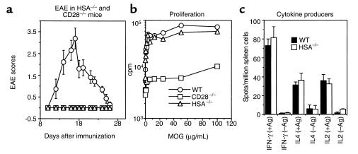
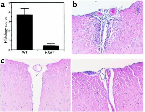
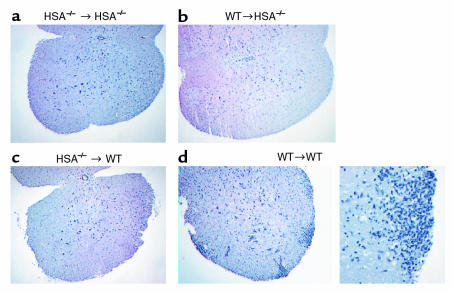
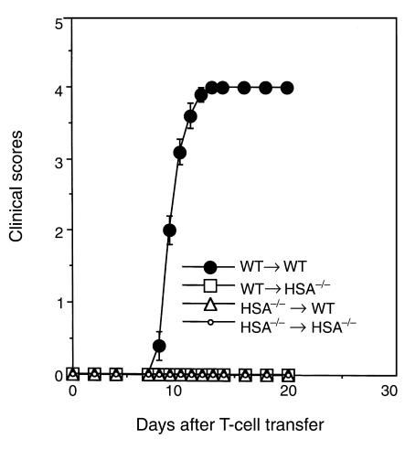
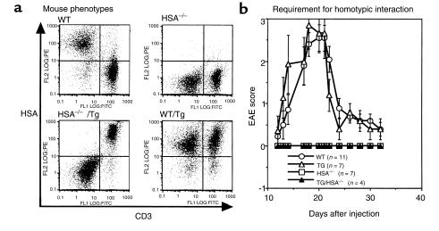
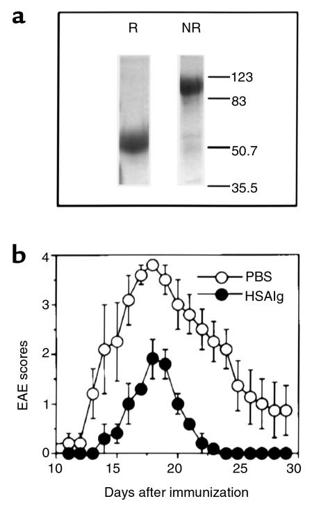
Induction of myelin-specific CD4 T cells is a pivotal event in the development of experimental autoimmune encephalomyelitis (EAE). Other checkpoints in EAE pathogenesis have not been clearly defined, although multiple genetic loci are known to influence EAE development. We report here that targeted mutation of the heat-stable antigen (HSA) abrogates development of EAE despite a complete lack of effect on induction of autoimmune T cells. To test whether T-cell expression of HSA is sufficient, we created transgenic mice in which HSA is expressed exclusively in the T-cell lineage. We found that these mice remain resistant to EAE induction. Adoptive transfer studies demonstrate that both T cells and non-T cells must express HSA in order for the pathogenic T cells to execute their effector function. Moreover, HSAIg, a fusion protein consisting of the extracellular domain of the HSA and the Fc portion of immunoglobulin, drastically ameliorates the clinical sign of EAE even when administrated after self-reactive T cells had been expanded. Thus, identification of HSA as a novel checkpoint, even after activation and expansion of self-reactive T cells, provides a novel approach for immunotherapy of autoimmune neurologic diseases, such as multiple sclerosis.


Figures

Figure 1
Figure 1
Targeted mutations of HSA and CD28 reveal two distinct checkpoints in the development of EAE. (a) Targeted mutations of either HSA or CD28 prevent induction of EAE. WT, CD28–/–, or HSA–/– mice were immunized with MOG peptide. Clinical signs were scored as described in Methods. (b) Proliferative response of lymph node T cells to MOG peptides. Draining lymph node cells from day 10–immunized mice were stimulated with given concentrations of MOG peptide and irradiated syngeneic naive spleen cells as antigen-presenting cells. (c) Enumeration of cytokine-producers by ELISpot. Draining lymph node cells used in b were used as responder cells. The numbers of cells secreting either IL2, IL4, or IFN-γ among 1 × 10⁶ lymph node cells in response to MOG peptide (AA35-55) are presented. Data shown are means ± SEM from three independent experiments.
Figure 2
Figure 2
Histological analysis of spinal cord of MOG-immunized WT or HSA–/– mice. (a) The means and SEM of histological scores of WT and HSA–/– mice spinal cords. Ten independent cross sections, from cervical to sacral regions, were examined in each spinal cord. The data are summarized from 30 spinal cord sections from three mice in each group. (b) Representative histology in immunized WT mice; all sections examined contain histology lesions. (c) Histology sections (×100) of immunized HSA–/– mice. A lesion-free section is presented on the left, and a lesion-containing section is presented on the right.
Figure 3
Figure 3
Requirement for HSA expression on both T cells and non-T host cells for the induction of EAE. Histology (×63 for ac and the left side of d; ×200 for the right side of d) of spinal cords of the HSA–/– (a and b) or WT (c and d) recipient mice on day 12 after adoptive transfer. Draining lymph node cells were isolated from either WT or HSA–/– mice after immunization and were stimulated with antigen and IL-2 for 4 days in vitro. The activated T cells were injected into either WT or HSA–/– mice (108 cells per mouse). EAE development was monitored daily for clinical signs. At 12 days after transfer, recipient mice were sacrificed and spinal cords were processed for histological examination. No disease was observed in WT → HSA–/–, HSA–/– → WT, or HSA–/– → HSA–/– recipients.
Figure 4
Figure 4
Clinical scores of the adoptive transfer experiment with four (WT → HSA–/– and HSA–/– → WT groups) or five (WT → WT and HSA–/– → HSA–/– groups) mice per group.
Figure 5
Figure 5
Transgenic expression of HSA exclusively on T-cell lineage is insufficient for EAE development. (a) Phenotypes of WT, HSA-TG, HSA–/–, and HSATG/HSA–/– mice by flow cytometry using anti-HSA and anti-CD3 mAb’s. (b) EAE score in WT, HSA-TG, HSA–/–, and HSATG/HSA–/– mice after immunization with the MOG peptides.
Figure 6
Figure 6
HSAIg ameliorates EAE. (a) Analysis of HSAIg by SDS-PAGE. A total of 10 μg of purified HSAIg was separated by 10% reducing (R) and nonreducing (NR) SDS-PAGE. The proteins were stained by Coomassie blue. (b) The EAE score for control (PBS) or HSAIg-treated mice. EAE was induced in WT mice as described in Methods. On days 8, 10, 12, 14, and 22 after immunization, five mice per group were injected intraperitoneally with 100 μg/mouse of either HSAIg or 100 μL of PBS as control. The effect of HSAIg has been evaluated in three independent experiments with similar results.

References

    1. Mendel I, Kerlero de Rosbo N, Ben-Nun A. A myelin oligodendrocyte glycoprotein peptide induces typical chronic experimental autoimmune encephalomyelitis in H-2b mice: fine specificity and T cell receptor V beta expression of encephalitogenic T cells. Eur J Immunol. 1995;25:1951–1959. - PubMed
    1. Kerlero de Rosbo N, Mendel I, Ben-Nun A. Chronic relapsing experimental autoimmune encephalomyelitis with a delayed onset and an atypical clinical course, induced in PL/J mice by myelin oligodendrocyte glycoprotein (MOG)-derived peptide: preliminary analysis of MOG T cell epitopes. Eur J Immunol. 1995;25:985–993. - PubMed
    1. Zamvil SS, Steinman L. The T lymphocyte in experimental allergic encephalomyelitis. Annu Rev Immunol. 1990;8:579–621. - PubMed
    1. Baron JL, Madri JA, Ruddle NH, Hashim G, Janeway CA., Jr Surface expression of alpha 4 integrin by CD4 T cells is required for their entry into brain parenchyma. J Exp Med. 1993;177:57–68. - PMC - PubMed
    1. Vanderlugt CL, et al. The functional significance of epitope spreading and its regulation by co-stimulatory molecules. Immunol Rev. 1998;164:63–72. - PubMed

Publication types

MeSH terms