. 2000 May;105(9):1233-41.
doi: 10.1172/JCI7610.

Epidermal growth factor receptor expression in neurofibromatosis type 1-related tumors and NF1 animal models

Affiliations

Epidermal growth factor receptor expression in neurofibromatosis type 1-related tumors and NF1 animal models

J E DeClue et al. J Clin Invest. 2000 May.

Abstract

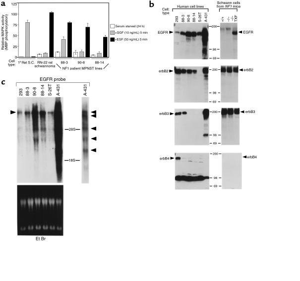
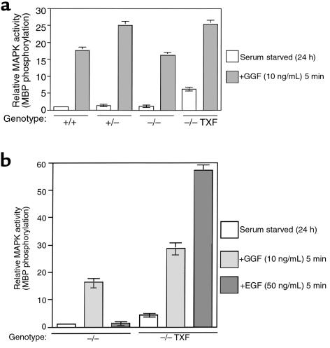
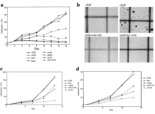
We have found that EGF-R expression is associated with the development of the Schwann cell-derived tumors characteristic of neurofibromatosis type 1 (NF1) and in animal models of this disease. This is surprising, because Schwann cells normally lack EGF-R and respond to ligands other than EGF. Nevertheless, immunoblotting, Northern analysis, and immunohistochemistry revealed that each of 3 malignant peripheral nerve sheath tumor (MPNST) cell lines from NF1 patients expressed the EGF-R, as did 7 of 7 other primary MPNSTs, a non-NF1 MPNST cell line, and the S100(+) cells from each of 9 benign neurofibromas. Furthermore, transformed derivatives of Schwann cells from NF1(-/-) mouse embryos also expressed the EGF-R. All of the cells or cell lines expressing EGF-R responded to EGF by activation of downstream signaling pathways. Thus, EGF-R expression may play an important role in NF1 tumorigenesis and Schwann cell transformation. Consistent with this hypothesis, growth of NF1 MPNST lines and the transformed NF1(-/-) mouse embryo Schwann cells was greatly stimulated by EGF in vitro and could be blocked by agents that antagonize EGF-R function.


Figures

Figure 1
Figure 1
Response of primary rat Schwann cells, RN-22 rat schwannoma line, and human NF1 patient MPNST lines to GGF and EGF, and expression of EGF-R and erbB2, -3, and -4 proteins and EGFR mRNA. (a) The indicated cells were grown until nearly confluent, serum starved for 24 hours, then left untreated (–) or stimulated with 10 ng/mL recombinant human GGF (G) or 50 ng/mL recombinant human EGF (E) for 5 minutes at 37°C. The cells were lysed and the endogenous MAP kinase activity was assayed. Following the reaction, incorporation of 32Pi into exogenous myelin basic protein was determined. Values were normalized to unstimulated primary rat Schwann cells (1.0) and represent the results of two experiments, carried out in duplicate, with error bars shown. (b) Expression of EGF-R and erbB2, -3, and -4 proteins in human and mouse cells. Cells were grown until confluent and lysed, and lysates containing 50 μg human or 100 μg mouse cell protein were subjected to analysis by SDS-PAGE and immunoblotting using antibodies specific for each protein indicated (arrows). 293, human embryonic kidney cells; 88-3, 90-8, 88-14, human NF1 MPNST lines; S-26T, human non-NF1 MPNST line; A-431, human epidermoid carcinoma line. Migration of molecular standards (kDa) is indicated at center. A strong nonspecific band of approximately 90 kDa appeared in the erbB4 blot of human but not mouse lysates. (c) Expression of EGFR mRNA in human MPNST and control cell lines. Cells were grown until confluent and lysed, and 20 μg of total RNA from each line was subjected to electrophoresis, transferred to a filter, and hybridized to a human EGFR probe labeled with 32P. The predominant 10.5-kb mRNA is indicated with an arrow at left, as is the approximate location of the 28S and 18S RNAs (top). At right is a shorter exposure of the A-431 line, with arrows designating the different mRNAs detected. The filter was photographed under ultraviolet light before hybridization (bottom).
Figure 2
Figure 2
EGF-R expression in NF1 tumor sections. Paraffin sections were stained with anti–EGF-R (b, d, and f) or anti-S100 (a, c, and e) or with both antibodies (g, h, and i). Visualization of single antibodies was with 3′3-diaminobenzidine HCl (DAB; brown) (af). Double labeling used nitroblue tetrazolium (NBT/BCIP; blue) for anti-S100, and DAB (brown) for anti–EGF-R (gi). In a, b, and df, the counterstain is hematoxylin (blue); in c and gi, the counterstain is nuclear fast red (pink). (ad) Sections from MPNST. In a, the arrow points to a normal nerve within the tumor, containing S100+ cells, whereas the tumor matrix is S100. An adjacent section from the same tumor in b shows that most cells are EGF-R+. Another MPNST contains S100+ cells (c) and is mostly negative for EGF-R (d). (e and f) Sections of normal human nerve (arrow points to perineurium). (g) A section of a cutaneous neurofibroma with EGF-R+/S100+ cells. (h) A section from a dissociated neurofibroma cell preparation. The arrow designates a group of cells double stained by anti-S100 and anti–EGF-R. (i) A section from a dissociated normal nerve preparation. No double-labeled cells are detected. ag, bar: 34.2 μm; h and i, bar: 16.3 μm.
Figure 3
Figure 3
Transformed derivatives of NF1 (–/–) mouse embryo–derived Schwann cells have elevated basal MAP kinase activity and respond to EGF. (a) Mouse Schwann cells were isolated from day 12.5 embryos with wild-type NF1 (+/+) or targeted disruption of one NF1 allele (+/–), or both alleles (–/–), and compared with transformed derivatives of (–/–) (TXF). The cells were serum starved for 24 hours, then left untreated or stimulated with GGF, as indicated. Cells were lysed and MAP kinase activity was determined as for Figure 1. Results are the mean of two experiments carried out in duplicate, with error bars shown. Results were normalized to the level of activity present in serum-starved wild-type (+/+) cells (1.0). (b) Schwann cells from (–/–) embryos and TXF were treated and assayed as above, except additional samples were prepared after stimulation with 50 ng/mL EGF for 5 minutes. Results are the mean of two experiments carried out in duplicate, with error bars shown. Results were normalized to the level of activity present in serum-starved homozygous null (–/–) cells (1.0).
Figure 4
Figure 4
Growth of NF1 patient MPNST lines is EGF dependent and is inhibited by EGF-R antagonists. (a) MPNST line 88-14 was plated at 2 × 10⁴ per 35-mm well. The next day (day 1) cells were switched to medium without (–EGF) or with 10 ng/mL EGF (+EGF), and on day 2 cells were switched to medium containing 10 ng/mL EGF and 3% PBS (Veh1); 0.1% DMSO (Veh2); 3 μg/mL mAb 225 in PBS (3% vol/vol; mAb); 40 μM farnesyltransferase inhibitor B581 in water; 10 μM tyrphostin A-25 in DMSO; or 400 nM tyrphostin AG-1478, also in DMSO. Cells were re-fed with medium containing fresh inhibitor and counted in duplicate every 2 days, beginning at day 2. (b) Growth of MPNST 88-14 line in soft agar. Cells were plated at 105/mL in a 0.4% (wt/vol) agar suspension with 7% FBS with or without 100 ng/mL EGF, as indicated; mAb 225 was included at 5 μg/mL and AG-1478 at 400 nM. Cells were photographed after 4 weeks. × 25. (c and d) Growth of 88-14 (c) and 90-8 (d) in 2% serum is inhibited by EGF-R antagonists. Cells were plated as in a, and switched on day 1 to medium containing 2% FBS (CON) or 2% serum plus 0.1% DMSO (Veh); 3 μg/mL mAb 225 (mAb); 10 μM tyrphostin A-25; or 400 nM tyrphostin AG-1478. Thereafter, cells were re-fed with medium containing fresh inhibitor every 2 days, and duplicate wells of cells were counted every three days.

References

    1. Huson SM, Harper PS, Compston D. Von Recklinghausen neurofibromatosis: a clinical and population study in south-east Wales. Brain. 1988;111:1355–1381. - PubMed
    1. Upadhyaya, M., and Cooper, D.N. 1998. Neurofibromatosis type 1: from genotype to phenotype. BIOS Scientific Publishers Ltd. Oxford, United Kingdom. pp. 230.
    1. Ferner, R.E. 1998. Clinical aspects of neurofibromatosis 1. In Neurofibromatosis type 1: from genotype to phenotype. M. Upadhyaya and D.N. Cooper, editors. BIOS Scientific Publishers Ltd., Oxford, United Kingdom. 21–38.
    1. Rosenbaum T, Petrie KM, Ratner N. Neurofibromatosis type 1: genetic and cellular mechanisms of peripheral nerve tumor formation. The Neuroscientist. 1997;3:412–420.
    1. Stefansson K, Wollmann R, Jerkovic M. S-100 protein in soft tissue tumours derived from Schwann cells and melanocytes. Am J Pathol. 1982;106:261–268. - PMC - PubMed

Publication types