. 2000 May;105(9):1269-77.
doi: 10.1172/JCI5250.

Corticotropin-releasing hormone links pituitary adrenocorticotropin gene expression and release during adrenal insufficiency

Affiliations

Corticotropin-releasing hormone links pituitary adrenocorticotropin gene expression and release during adrenal insufficiency

L J Muglia et al. J Clin Invest. 2000 May.

Abstract

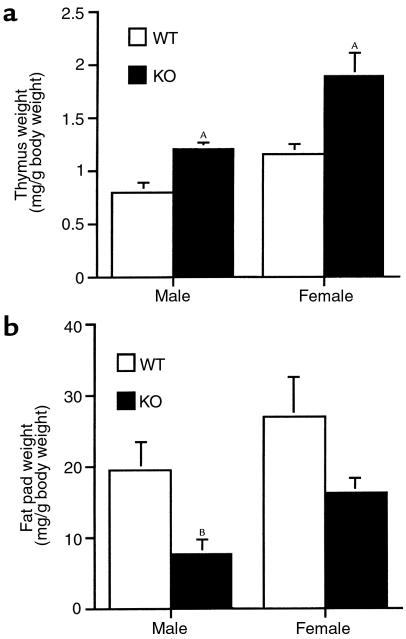
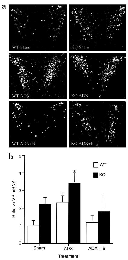
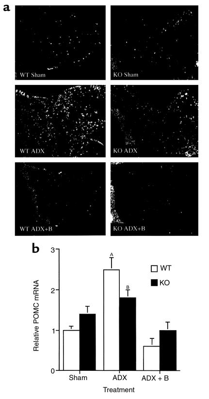
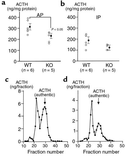
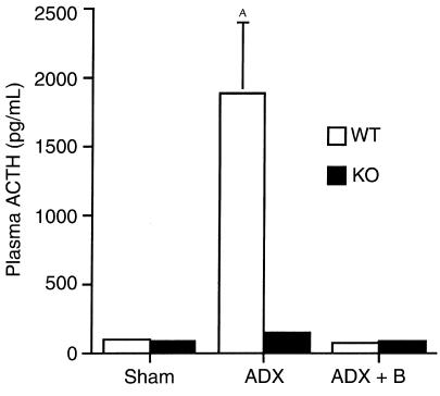
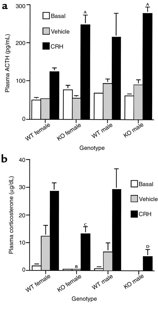
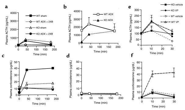
Corticotropin-releasing hormone (CRH)-deficient (KO) mice provide a unique system to define the role of CRH in regulation of the hypothalamic-pituitary-adrenal (HPA) axis. Despite several manifestations of chronic glucocorticoid insufficiency, basal pituitary proopiomelanocortin (POMC) mRNA, adrenocorticotrophic hormone (ACTH) peptide content within the pituitary, and plasma ACTH concentrations are not elevated in CRH KO mice. The normal POMC mRNA content in KO mice is dependent upon residual glucocorticoid secretion, as it increases in both KO and WT mice after adrenalectomy; this increase is reversed by glucocorticoid, but not aldosterone, replacement. However, the normal plasma levels of ACTH in CRH KO mice are not dependent upon residual glucocorticoid secretion, because, after adrenalectomy, these levels do not undergo the normal increase seen in KO mice despite the increase in POMC mRNA content. Administration of CRH restores ACTH secretion to its expected high level in adrenalectomized CRH KO mice. Thus, in adrenal insufficiency, loss of glucocorticoid feedback by itself can increase POMC gene expression in the pituitary; but CRH action is essential for this to result in increased secretion of ACTH. This may explain why, after withdrawal of chronic glucocorticoid treatment, reactivation of CRH secretion is a necessary prerequisite for recovery from suppression of the HPA axis.


Figures

Figure 1
Figure 1
Evidence of chronic glucocorticoid insufficiency in CRH-deficient mice. (a) Thymus weight normalized to total body weight for WT and CRH-deficient (KO) mice of each sex. AP < 0.01 vs. WT of same sex. (b) Testicular (male) or ovarian (female) fat pad weight normalized to total body weight. BP < 0.05 vs. WT male.
Figure 2
Figure 2
Effect of adrenalectomy (ADX) and low-dose corticosterone replacement on hypothalamic VP mRNA in WT and CRH KO mice. (a) Representative panels (from n = 3 per genotype and treatment group) of emulsion-dipped coronal sections through the hypothalamic paraventricular nucleus subjected to in situ hybridization with a radiolabeled VP antisense riboprobe. Basal VP mRNA tended to be elevated in KO sham as compared with WT mice, and was further induced after adrenalectomy. VP mRNA was suppressed in WT and KO mice given low-dose corticosterone after adrenalectomy (ADX + B). (b) Quantitative analysis of VP mRNA hybridization signal intensity in the paraventricular nucleus. Pairwise comparisons: borderline significant increase WT ADX vs. WT sham (AP = 0.06); BP < 0.05 CRH KO ADX vs. WT sham.
Figure 3
Figure 3
Effect of adrenalectomy (ADX) and low-dose corticosterone replacement on AP POMC mRNA in WT and CRH KO mice. (a) In situ hybridization of the pituitary with a radiolabeled POMC antisense riboprobe (representative of n = 3 per genotype and treatment group). The neurointermediate lobe is oriented at the left side of each panel. Basal levels of POMC mRNA are equivalent in WT and KO mice, and increase after adrenalectomy in AP corticotrophs. The neurointermediate lobe of the pituitary demonstrated unchanged hybridization intensity before and after adrenalectomy. (b) Quantitative analysis of POMC mRNA hybridization signal intensity in the AP. AP = 0.01 vs. WT Sham and WT ADX+B; BP < 0.05 vs. WT Sham and WT ADX + B.
Figure 4
Figure 4
Pituitary ACTH content and processing in CRH KO and WT mice. Immunoreactive ACTH contents of (a) anterior pituitary (AP) and (b) intermediate-posterior pituitary (IP) from individual mice and means ± SEM are shown. AP < 0.05 vs. WT AP. Representative elution profiles of Sephadex G-50 gel filtration chromatography of ACTH extracted from AP of (c) WT and (d) CRH KO mice.
Figure 5
Figure 5
Effect of adrenalectomy (ADX) and low-dose corticosterone replacement (ADX + B) on plasma ACTH in WT and CRH KO mice. Plasma ACTH increases to a much greater extent in WT than KO mice despite similar induction of pituitary POMC mRNA. Low-level glucocorticoid supplementation after adrenalectomy effectively returns plasma ACTH to basal levels. AP < 0.05 vs. all other groups.
Figure 6
Figure 6
CRH stimulation of WT and CRH KO mice. (a) Plasma ACTH concentration 30 minutes after either no injection (basal), or intraperitoneal injection of vehicle or 10 μg/kg CRH. AP < 0.005 vs. WT female CRH. (b) Plasma corticosterone concentration after CRH, vehicle, or no injection. BP < 0.05 vs. WT female vehicle; CP < 0.01 vs. WT female CRH; DP < 0.05 vs. WT male CRH.
Figure 7
Figure 7
Stimulation of the HPA axis by CRH (ad) or vasopressin (e and f) in WT and CRH KO mice. Time course for plasma ACTH is shown for sham-adrenalectomized (sham) or adrenalectomized mice supplemented with constant-release corticosterone pellets delivering double the physiologic replacement (ADX + 2XB) (a), and for adrenalectomized (ADX) mice (b), after intraperitoneal injection of 90 μg/kg CRH. Respective time courses for plasma corticosterone are shown for the mice in a (c) and b (d). Plasma ACTH (e) and corticosterone (f) after injection of vehicle or VP. Plasma ACTH increases to a similar extent in KO and WT mice after VP compared with vehicle injection, although only the WT mice demonstrate a significant increase in plasma corticosterone.

Comment in

References

    1. Chrousos GP. The hypothalamic-pituitary-adrenal axis and immune-mediated inflammation. N Engl J Med. 1995;332:1351–1362. - PubMed
    1. Munck A, Guyre PM, Holbrook NJ. Physiological functions of glucocorticoids in stress and their relation to pharmacological actions. Endocr Rev. 1984;5:25–44. - PubMed
    1. Orth, D.N., Kovacs, W.J., and Debold, C.R. 1992. The adrenal cortex. In Williams textbook of endocrinology. J.D. Wilson and D.W. Foster, editors. W.B. Saunders Co. Philadelphia, Pennsylvania, USA. 489–619.
    1. Suda T, et al. The role of corticotropin-releasing factor and vasopressin in hypoglycemia induced proopiomelanocortin gene expression in the rat anterior pituitary gland. Brain Res. 1992;579:303–308. - PubMed
    1. Plotsky PM, Bruhn TO, Vale W. Hypophysiotropic regulation of adrenocorticotropin secretion in response to insulin-induced hypoglycemia. Endocrinology. 1985;117:323–329. - PubMed

Publication types